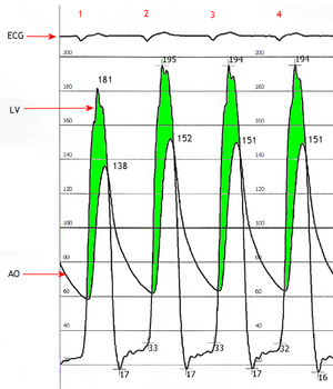
Simultaneous left ventricular and aortic pressure tracings demonstrate a pressure gradient between the left ventricle and aorta, suggesting aortic stenosis. The left ventricle generates higher pressures than what is transmitted to the aorta.  The pressure gradient, caused by aortic stenosis, is represented by the green shaded area. (AO = ascending aorta; LV = left ventricle; ECG = electrocardiogram.)
Find information on thousands of medical conditions and prescription drugs.

Aortic valve stenosis

Aortic valve stenosis (AS) is a heart condition caused by the incomplete opening of the aortic valve. more...

Home
Diseases
A
Aagenaes syndrome
Aarskog Ose Pande syndrome
Aarskog syndrome
Aase Smith syndrome
Aase syndrome
ABCD syndrome
Abdallat Davis Farrage...
Abdominal aortic aneurysm
Abdominal cystic...
Abdominal defects
Ablutophobia
Absence of Gluteal muscle
Acalvaria
Acanthocheilonemiasis
Acanthocytosis
Acarophobia
Acatalasemia
Accessory pancreas
Achalasia
Achard syndrome
Achard-Thiers syndrome
Acheiropodia
Achondrogenesis
Achondrogenesis type 1A
Achondrogenesis type 1B
Achondroplasia
Achondroplastic dwarfism
Achromatopsia
Acid maltase deficiency
Ackerman syndrome
Acne
Acne rosacea
Acoustic neuroma
Acquired ichthyosis
Acquired syphilis
Acrofacial dysostosis,...
Acromegaly
Acrophobia
Acrospiroma
Actinomycosis
Activated protein C...
Acute febrile...
Acute intermittent porphyria
Acute lymphoblastic leukemia
Acute lymphocytic leukemia
Acute mountain sickness
Acute myelocytic leukemia
Acute myelogenous leukemia
Acute necrotizing...
Acute promyelocytic leukemia
Acute renal failure
Acute respiratory...
Acute tubular necrosis
Adams Nance syndrome
Adams-Oliver syndrome
Addison's disease
Adducted thumb syndrome...
Adenoid cystic carcinoma
Adenoma
Adenomyosis
Adenosine deaminase...
Adenosine monophosphate...
Adie syndrome
Adrenal incidentaloma
Adrenal insufficiency
Adrenocortical carcinoma
Adrenogenital syndrome
Adrenoleukodystrophy
Aerophobia
Agoraphobia
Agrizoophobia
Agyrophobia
Aicardi syndrome
Aichmophobia
AIDS
AIDS Dementia Complex
Ainhum
Albinism
Albright's hereditary...
Albuminurophobia
Alcaptonuria
Alcohol fetopathy
Alcoholic hepatitis
Alcoholic liver cirrhosis
Alektorophobia
Alexander disease
Alien hand syndrome
Alkaptonuria
Alliumphobia
Alopecia
Alopecia areata
Alopecia totalis
Alopecia universalis
Alpers disease
Alpha 1-antitrypsin...
Alpha-mannosidosis
Alport syndrome
Alternating hemiplegia
Alzheimer's disease
Amaurosis
Amblyopia
Ambras syndrome
Amelogenesis imperfecta
Amenorrhea
American trypanosomiasis
Amoebiasis
Amyloidosis
Amyotrophic lateral...
Anaphylaxis
Androgen insensitivity...
Anemia
Anemia, Diamond-Blackfan
Anemia, Pernicious
Anemia, Sideroblastic
Anemophobia
Anencephaly
Aneurysm
Aneurysm
Aneurysm of sinus of...
Angelman syndrome
Anguillulosis
Aniridia
Anisakiasis
Ankylosing spondylitis
Ankylostomiasis
Annular pancreas
Anorchidism
Anorexia nervosa
Anosmia
Anotia
Anthophobia
Anthrax disease
Antiphospholipid syndrome
Antisocial personality...
Antithrombin deficiency,...
Anton's syndrome
Aortic aneurysm
Aortic coarctation
Aortic dissection
Aortic valve stenosis
Apert syndrome
Aphthous stomatitis
Apiphobia
Aplastic anemia
Appendicitis
Apraxia
Arachnoiditis
Argininosuccinate...
Argininosuccinic aciduria
Argyria
Arnold-Chiari malformation
Arrhythmogenic right...
Arteriovenous malformation
Arteritis
Arthritis
Arthritis, Juvenile
Arthrogryposis
Arthrogryposis multiplex...
Asbestosis
Ascariasis
Aseptic meningitis
Asherman's syndrome
Aspartylglycosaminuria
Aspergillosis
Asphyxia neonatorum
Asthenia
Asthenia
Asthenophobia
Asthma
Astrocytoma
Ataxia telangiectasia
Atelectasis
Atelosteogenesis, type II
Atherosclerosis
Athetosis
Atopic Dermatitis
Atrial septal defect
Atrioventricular septal...
Atrophy
Attention Deficit...
Autoimmune hepatitis
Autoimmune...
Automysophobia
Autonomic dysfunction
Familial Alzheimer disease
Senescence
B
C
D
E
F
G
H
I
J
K
L
M
N
O
P
Q
R
S
T
U
V
W
X
Y
Z
Medicines

The aortic valve controls the direction of blood flow from the left ventricle to the aorta. When in good working order, the aortic valve does not impede the flow of blood between these two spaces. Under some circumstances, the aortic valve becomes narrower than normal, impeding the flow of blood. This is known as aortic valve stenosis, or aortic stenosis, often abbreviated as AS.

Pathophysiology

When the aortic valve becomes stenotic, it causes a pressure gradient between the left ventricle (LV) and the aorta. The more constricted the valve, the higher the gradient between the LV and the aorta. For instance, with a mild AS, the gradient may be 20 mmHg. This means that, at peak systole, while the LV may generate a pressure of 140 mmHg, the pressure that is transmitted to the aorta will only be 120 mmHg. So, while a blood pressure cuff may measure a normal systolic blood pressure, the actual pressure generated by the LV would be considerably higher.

In individuals with AS, the left ventricle (LV) has to generate an increased pressure in order to overcome the increased afterload caused by the stenotic aortic valve and eject blood out of the LV. The more severe the aortic stenosis, the higher the gradient is between the left ventricular systolic pressures and the aortic systolic pressures. Due to the increased pressures generated by the left ventricle, the myocardium (muscle) of the LV undergoes hypertrophy (increase in muscle mass). This is seen as thickening of the walls of the LV. The type of hypertrophy most commonly seen in AS is concentric hypertrophy, meaning that all the walls of the LV are (approximately) equally thickened.

Etiology

Causes of aortic stenosis include acute rheumatic fever, bicuspid aortic valve and congenital anomalies. As individuals age, calcification of the aortic valves may occur and result in stenosis.

Physical examination

It is most often diagnosed when it is asymptomatic. It is found on routine examination of the heart. A fairly loud systolic, crescendo-decrescendo murmur is heard loudest at the upper right sternal border, and radiates to the carotid arteries. The murmur increases with squatting, decreases with standing and isometric muscular contraction, which helps distinguish it from hypertrophic obstructive cardiomyopathy (HOCM). Respiration has no effect on the loudness of the murmur. The more severe the degree of the stenosis, the later the peak occurs in the crescendo-decrescendo of the murmur. Due to increases in left ventricular pressure from the stenotic aortic valve, over time the ventricle may hypertrophy, resulting in a diastolic dysfunction. As a result, one may hear a 4th heart sound due to the stiff ventricle. With continued increases in ventricular pressure, dilatation of the ventricle will occur, and a 3rd heart sound may be manifest.

Read more at Wikipedia.org


[List your site here Free!]


Aortic valve stenosis
From Gale Encyclopedia of Medicine, 4/6/01 by Jeanine Barone

Definition

When aortic valve stenosis occurs, the aortic valve, located between the aorta and left ventricle of the heart, is narrower than normal size.

Description

A normal aortic valve, when open, allows the free flow of blood from the left ventricle to the aorta. When the valve narrows, as it does with stenosis, blood flow is impeded. Because it is more difficult for blood to flow through the valve, there is increased strain on the heart. This can cause the left ventricle to enlarge and malfunction, resulting in reduced blood supply to the heart muscle and body, as well as fluid build up in the lungs.

Cause & symptoms

Aortic valve stenosis can occur because of a birth defect in the formation of the valve. Calcium deposits may form on the valve with aging, causing the valve to become stiff and narrow. Stenosis can also occur as a result of rheumatic fever. Mild aortic stenosis may produce no symptoms at all. The most common symptoms, depending on the severity of the disease, are chest pain, blackouts, and difficulty breathing.

Diagnosis

Using a stethoscope, a physician may hear a murmur and other abnormal heart sounds. An ECG, also called an electrocardiogram, records the electrical activity of the heart. This technique and chest x ray can show evidence that the left ventricle is enlarged. An x ray can also reveal calcium deposits on the valve, as well as congestion in the lungs. Echocardiography can pick up thickening of the valve, heart size, and whether or not the valve is working properly. This is a procedure in which high frequency sound waves harmlessly bounce off organs in the body. Cardiac catheterization, in which a contrast dye is injected in an artery using a catheter, is the key tool to confirm stenosis and gauge its severity.

Treatment

Treatment depends on the symptoms and how the heart's function is affected. The valve can be opened without surgery by using a balloon catheter, but this is often a temporary solution. The procedure involves inserting a deflated balloon at the end of a catheter through the arteries to the valve. Inflating the balloon should widen the valve. In severe stenosis, heart valve replacement is recommended, most often involving open-heart surgery. The valve can be replaced with a mechanical valve, a valve from a pig, or by moving the patient's other heart valve (pulmonary) into the position of the aortic valve and then replacing the pulmonary valve with an mechanical one. Anyone with aortic stenosis needs to take antibiotics (amoxicillin, erythromycin, or clindamycin) before dental and some other surgical procedures, to prevent a heart valve infection.

Prognosis

The prognosis for aortic valve stenosis depends on the severity of the disease. With surgical repair, the disease is curable. Patients suffering mild stenosis can usually lead a normal life; a minority of the patients progress to severe disease. Anyone with moderate stenosis should avoid vigorous physical activity. Most of these patients end up suffering some kind of coronary heart disease over a 10 year period. Because it is a progressive disease, moderate and severe stenosis will be treated ultimately with surgery. Severe disease, if left untreated, leads to death within 2 to 4 years once the symptoms start.

Prevention

There is no way to prevent aortic stenosis.

Key Terms

Aorta
The largest artery in the body, which moves blood from the left ventricle to the rest of the body.
ECG
Also called an electrocardiogram, it records the electrical activity of the heart.
Echocardiogram
A procedure in which high frequency sound waves harmlessly bounce off organs in the body providing an image so one can determine their structure and function.
Cardiac catheterization
A procedure in which dye is injected through a tube or catheter into an artery to more easily observe valves or blood vessels seen on an x ray.
Left ventricle
One of the lower chambers of the heart, which pumps blood to the aorta.
Murmur
An abnormal heart sound that can reflect a valve dysfunction.
Rheumatic fever
A bacterial infection that often causes heart inflammation.
Pulmonary valve
The valve located between the pulmonary artery and the right ventricle, which brings blood to the lungs.

Further Reading

For Your Information

    Books

  • Bender, Jeffrey R. "Heart Valve Disease." In Yale University School of Medicine Heart Book, edited by Barry L. Zaret, et al. New York: Hearst Books, 1992, 167-175.
  • Braunwald, E. "Aortic Stenosis." In Heart Disease: A Textbook of Cardiovascular Medicine. Philadelphia: W.B. Saunders Company, 1997, 1035-1045.
  • McGoon, Michael D. "Aortic Stenosis." In Mayo Clinic Heart Book. New York: William Morrow and Company, 1993, 65-66.

    Other

  • Loyola University Medical Center. "Aortic Stenosis," by Aly Rahimtoola. http://www.meddean.luc.edu/develop/arahimtoola/intro.htm.
  • Ochsner Heart and Vascular Institute. "Aortic Stenosis." http://www.ochsner.org/pedcard/as.htm.

Gale Encyclopedia of Medicine. Gale Research, 1999.

Return to Aortic valve stenosis
Home Contact Resources Exchange Links ebay