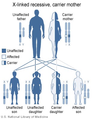
Myotonias are rare disorders characterized by difficulties in skeletal muscle relaxation. Either dominant or recessive modes of inheritance are possible. Underlying gene mutations cause defects in the ion channels of the muscle membranes. Previously undiagnosed myotonias may occur among military conscripts. We report here eight such patients with enhanced symptoms of myotonia during their military service. Six patients had myotonia congenita, one had myotonic dystrophy, and one paramyotonia congenita. In myotonia congenita, serum creatine kinase and aldolase levels correlated with the recommended service fitness classification. Because some anesthetic agents may have unfavorable side effects in myotonia, both patients and anesthesiologists need to be aware of the diagnosis. The awareness of military surgeons regarding the possibility of myotonia is necessary to provide a correct diagnosis and to establish the service fitness of these patients.
Introduction
Myotonia means difficulty in the relaxation of skeletal muscle after voluntary contraction. It is present with initial activation and usually abates after repeated muscle activity (warm-up phenomenon). The diagnosis is based on a careful neurological examination demonstrating myotonic features of muscles and, usually, skeletal muscle hypertrophy. Electroneuronomyography (ENMG) reveals typical repetitive discharges of varying amplitude and frequency.1 There is considerable overlap between the various clinical subtypes, and the severity of the clinical findings varies considerably.2 Muscle biopsy findings are nonspecific in differential diagnoses of myotonias.1 Several gene defects affecting ion channels in muscle membranes may lead to myotonia. Direct identifications of gene mutations causing the disorder are frequently available for further classification.3 Myotonia occurs in several specific disorders; three of them, pertinent to this study, are described below (for a more extensive description, see, for example, Online Mendelian Inheritance in Man, at http://www.ncbi.nlm.nih.gov/omim).
In myotonia congenita, myotonia is most clearly demonstrated by an inability or delay in release of the hand grip. Muscle strength is usually normal at rest, but myotonia may cause functional difficulties in proximal muscles, for example while climbing stairs. Myotonia may lead to muscular hypertrophy, particularly in the limbs and trunk. Percussion myotonia, a local postpercussion contraction followed by abnormally slow muscle relaxation, occurs in approximately one-half of the patients. The symptoms are usually not aggravated by exposure to cold. Myotonia congenita is caused by mutations of the gene on chromosome 7 coding the proteins of the chloride channel of the skeletal muscle membrane. Both dominant (Thomsen's myotonia) and recessive (Becker's myotonia) modes of inheritance are possible.3
Paramyotonia congenita is a dominantly inherited myotonia aggravated by exercise and exposure to cold. It is often associated with muscle weakness or flaccid paresis after myotonia1 and with lability of serum potassium levels.4 The disease is nonprogressive; no atrophy or hypertrophy of the muscles is observed. Paramyotonia congenita results from mutations of a gene on chromosome 17 coding proteins of a natrium channel.3
Myotonic dystrophy is an autosomal dominant disorder characterized by myotonia and muscular dystrophy starting from the distal muscles of the extremities and the face, head, and neck. Cataracts, hypogonadism, frontal balding, and electrocardiographic changes usually occur as well. The symptoms typically manifest in middle age but may start during the second decade of life.1 A gene mutation on either chromosome 19 (type 1) or chromosome 3 (type 2) results in sodium and chloride channel defects. Cognitive performance declines in proportion to increased size of the gene defect and decreased age of onset of symptoms.5 Amplification of the gene defect is frequently observed after parent-to-child transmission.6
The drills during basic military training include exercises requiring rapid onset of movement from rest, without any possibility for individual stretching or warm-up. This may make myotonia particularly distinct. Moreover, myotonia is frequently increased by cold, hunger, fatigue, and emotional upset. All of these may occur during military service. Augmentation of myotonic symptoms during conscript service has been described.7
The prevalence of myotonia congenita in Northern Finland has recently been estimated to be 7.3 cases per 100,000. The disease affects male subjects more often than female subjects. The mean age of onset of symptoms is 11 years, but the mean time needed for diagnosis after the onset of symptoms is 18 years.8 Therefore, military conscripts may have undiagnosed myotonias. To facilitate detection of these patients, this report describes the symptoms and signs of myotonias diagnosed among conscripts and military regulars at Central Military Hospital during 1996-2002.
Patients and Findings
A retrospective review of the neurological medical records of Central Military Hospital of the Finnish Defense Forces for the years 1996-2002 revealed eight male patients (mean age, 22 years; range, 18-48 years) with previously undiagnosed myotonia. The mean age at the onset of symptoms was 9 years (range, 1-15 years). The duration of conscript service before the diagnosis was, on average, 2 months (range. 1-2.5 months). All patients were referred to the hospital by general practitioners working in garrisons. One of the authors (J.P.M.) examined all of the patients. A consulting clinical neurophysiologist performed ENMG.
For six patients (patients 1-6), clinical findings suggested myotonia congenita (Table I). One patient had myotonic dystrophy, and paramyotonia congenita was suspected for another. case reports present the pertinent findings for different diagnostic groups. ENMG revealed typical myotonic bursts from all limbs for all patients. The mean serum creatine kinase (CK) level for myotonia congenita patients was 270 U/L (range, 95-350 U/L; normal reference value,
Case Reports
Patient 1 had had stiffness of the lower limbs with sudden start of movements since the age of 8 years. The symptoms had spread to the upper limbs and neck and face muscles. After resting for 1 minute, new movements were associated with muscle cramps. In military service, the patient had fallen after being pushed slightly while running on stairs. During training in the field, his lower limbs stiffened after a jump over a ditch, and he fell into it. In an examination, strongly developed musculature was evident. The throat muscles stiffened after tongue protrusion. Release of the hand grip was slowed. Myotonia was also elicited from shoulder and thigh musculature. No percussion myotonia was observed. ENMG revealed myotonic discharges in muscles of all limbs. Myotonia congenita was diagnosed, and permanent exemption from military service was recommended.
Patient 5 was referred for neurological examination after ENMG, performed because of right shoulder pains, revealed myotonic discharges in all limbs, in addition to canalis carpi findings. The patient had previously had low back pain but had otherwise been healthy. Since the age of 10 years, he had had difficulties in starting fast movements and had easily stumbled in these instances. During wrestling, releasing hand grips had occasionally been difficult. During military service, the drill practices, particularly "falling out," had been difficult. Despite this, the patient graduated from the Finnish Military Academy, served as an artillery officer, obtained the rank of major, and was about to retire at the time of the examination. In brachioradialis reflex testing, the fingers returned to the rest position slowly, in a myotonic manner. A slightly prolonged release of the hand grip was also evident. Diadochokinesis and hand clapping were slightly slowed. No percussion myotonia was elicited. Myotonia congenita was diagnosed.
Patient 7 was referred for neurological examination because of slowness, clumsiness, and difficulties in finding him a post in the unit, 2 months after entering military service. In an examination, forehead wrinkling was slow. Release of the hand grip was delayed, and percussion myotonia was elicited from the trapezius muscle. Diadochokinesis and hand clapping were slowed. The CK level was 297 U/L and the aldolase level was 6.4 U/L. In a psychological examination, the general intelligence quotient was estimated to be 64 on the Wechsler Adult Intelligence Scale. In an ENMG examination, the patient showed a note indicating that his mother had been treated for myotonic dystrophy. ENMG revealed myotonic discharges. Myotonic dystrophy was diagnosed, and permanent exemption from military service was recommended.
Patient 8 had had difficulties with hand grip release 3 years before entering military service. His hands were sensitive to cold. His mother had suffered from muscle cramps. The patient's service began in January. During field service, his hands cramped for ~8 hours. During another field service episode 4 days later, the patient had hand cramps, shivering, and disorientation and was referred to a regional hospital. Deteriorated general condition and spastic upper limbs were observed; the serum potassium level was 2.7 mmol/L (normal reference range, 3.7-5.3 mmol/L) and the CK level was 630 U/L. Despite normal body temperature, C-reactive protein, and spinal tap findings, encephalitis was suspected, Acyclovir infusion was started and the patient was referred to Central Military Hospital for further treatment. On arrival, the patient was tired but coherent. Eye opening was difficult and muscle strength was decreased, although the patient was able to move all limbs. The muscles were painful. In brachioradialis reflex testing, the fingers returned to the rest position slowly, in a myotonic manner. ENMG revealed myotonic discharges; cold provocation produced muscle stiffness and pain but no additional ENMG features. During the follow-up period, serum potassium levels varied between 3.7 and 6.5 mmol/L; CK levels normalized to 121 U/L. Slow release of the hand grip was evident even at normal room temperature. Occasional neck stiffness occurred. The clinical picture agrees with paramyotonia congenita. Because of high personal motivation, service fitness class B and indoor service were recommended. The remaining period of conscript service was clinically uneventful.
Discussion
It is evident that previously undiagnosed myotonias occur among military conscripts. The condition should be suspected when the conscript complains about stiffness following fast movements after rest or displays unusual clumsiness, e.g., during drills. The duration of the hand grip release, possible percussion myotonia, and ENMG should be tested for these patients. The Finnish Defense Force's directions recommend exemption of patients with myotonic disorders from military service. However, variability of clinical expression is considerable, and individual evaluations may reveal suitable tasks for conscripts with high motivation. Myotonia congenita patients with the lowest CK and aldolase levels continued their military service (Table I). It appears that high muscle enzyme levels correlate with the severity of myotonic symptoms during service, possibly because of muscle injuries after small traumas or defects in the muscle membrane.
Symptoms of myotonia congenita may be alleviated by drugs that block voltage-dependent sodium channels, such as mexiletine and tocainide. Mexiletine is the drug of choice3 but has a narrow therapeutic range. Tocainide carries a risk of hematological problems. Drugs that raise the depolarization threshold of muscle membranes, such as procainamide, quinidine, phenytoin, or carbamazepine, are occasionally useful in myotonia as well.2,9 However, most patients with myotonia congenita cope well without medication.
Among patients with myotonia and paramyotonia congenita, anesthesia including suxamethonium to induce muscle relaxation may cause paradoxical muscle stiffness and render intubation or ventilation difficult. It is therefore important that the patients are aware of their disease, to alert the anesthesiologist about possible complications.2 In myotonic dystrophy, rehabilitation including maintenance of mobility and psychosocial support are necessary to optimize the possibilities of leading as normal a life as possible.
The impetus for publishing the first report on myotonia congenita by Thomsen in 1876 originated from the necessity of informing officials and colleagues, particularly military physicians, about the condition. Myotonia congenita ran in the family of Dr. Thomsen, and his youngest son, suffering from the same disease, was drafted by the Danish army. Despite letters from Thomsen and the mayor of their home city, his son was branded a malingerer, probably because of the son's well-developed musculature. After a 2-month period of military service, the officials were convinced about the disease and the son was exempted from service.10 It is worth noting that the 2-month observation period still occurs among patients with undiagnosed myotonias. case reports of myotonia patients describe an arrest attributable to disobedience for firing continuous bursts of shots instead of ordered single shots, because of the patient's inability to unclench his hand once having gripped the trigger of a machine gun on a cold morning.4
The symptoms of myotonia during military service are evident and the clinical signs easy to elicit. ENMG studies should be performed for all patients presenting such symptoms. In the future, additional diagnostic confirmation should be available from genetic testing, which also provides information on the risk of myotonia in the family and among offspring of the patients. The awareness of military surgeons about the possibility of myotonia is necessary to provide correct diagnosis and appropriate assessment of the fitness of these patients for military service.
Acknowledgment
Dr. M. Henriksson made valuable comments on this article.
References
1. Streib E: AAEE minimonograph #27: differential diagnosis of myotonic syndromes. Muscle Nerve 1987: 10: 603-15.
2. Russell S, Hirsch N: Anesthesia and myotonia. Br J Anesth 1994: 72: 210-6.
3. Kullmann D, Hanna M: Neurological disorders caused by inherited ion-channel mutations. Lancet Neurol 2002: 1: 157-66.
4. Hudson A: Progressive neurological disorder and myotonia associated with paramyotonia. Brain 1963: 86: 811-26.
5. Turnpenny P, Clark C. Kelly K: Intelligence quotient profile in myotonic dystrophy, intergenerational deficit, and correlation with CTG amplification. J Med Genet 1994; 31: 300-5.
6. Tsilfidis C. MacKenzie A, Mettler G, Barcelo J. Korneluk R: Correlation between CTG trinucleotide repeat length and frequency of severe congenital myotonic dystrophy. Nature Genet 1992: 1: 192-5.
7. Siirala M: Myotonia congenita (Thomsen's disease): clinical and pharmacological studies with special regard to the mechanism of the myotonic phenomenon. Ann AcadSciFenn 1949; 19: 1-117.
8. Baumann P. Myllyla V, Leisti J: Myotonia congenita in northern Finland: an epidemiologic and genetic study. J Med Genet 1998: 35: 293-6.
9. Sheela S: Myotonia congenita: response to carbamazepine. Ind Pediatr 2000: 37: 1122-5.
10. Hakulinen E: The man behind the syndrome: Julius Thomsen himself suffered of a type of myotonia [in Swedish}. Läkartidningen 1994; 91: 3178-80.
Guarantor: Jyrki P. Mäkelä, MD PhD
Contributors: Jyrki P. Mäkelä, MD PhD*[dagger]; Hannu Somer, MD PhD[double dagger]
* Central Military Hospital, P.O. Box 50, FIN-00301 Helsinki, Finland.
[dagger] BioMag Laboratory, Helsinki University Central Hospital, FIN-00290 Helsinki, Finland.
[double dagger] Department of Neurology, Helsinki University Central Hospital, FIN-00290 Helsinki, Finland.
This manuscript was received for review in February 2004 and was accepted for publication in August 2004.
Reprint & Copyright © by Association of Military Surgeons of U.S., 2005.
Copyright Association of Military Surgeons of the United States Sep 2005
Provided by ProQuest Information and Learning Company. All rights Reserved


