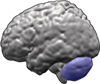
The relation between clinical outcome and single photon emission tomography (SPECT) results in cerebellitis has not been studied. A 63-year-old man developed cerebellar dysfunction with left emphasis one week after onset of cough. The only abnormality on analysis of cerebrospinal fluid was elevated protein (68 mg/dl). Magnetic resonance imaging was normal on the ninth day of ataxia. SPECT showed unilateral cerebellar hypoperfusion on the 13th day, but was normal on the 20th day. His gait improved a little by discharge on the 28th day and his tandem gait was only slightly unsteady six months later. This is the first evidence that normalization of cerebellar hypoperfusion in adult patients with cerebellitis is related to good outcome. Normalization of cerebellar hypoperfusion can occur in three weeks even when ataxia remains severe. [Neurol Res 2003; 25: 430-432]
Keywords: Cerebellitis; acute cerebellar ataxia; single photon emission tomography; hypoperfusion
INTRODUCTION
Cerebellitis, also referred to as acute cerebellar ataxia, is a neurological complication that may occur following viral infection. It is a relatively common condition in children and is considered a variant of post-infectious encephalomyelitis solely or predominantly involving the cerebellum. Magnetic resonance imaging (MRI) shows no abnormality in the majority of these cases1-4.
Only three reports in the English literature have focused on acute-stage findings of single photon emission tomography (SPECT) in cerebellitis1,2,5 . Both cerebellar hyperperfusion and hypoperfusion were reported. All patients were children except for one adolescent. The relation between serial follow-up SPECT and clinical outcome in cerebellitis has not been studied. Herein we report the first adult cerebellitis patient with rapid normalization of cerebellar hypoperfusion, which heralded slow but good functional recovery.
CASE REPORT
The patient was a 63-year-old married male, a retired teacher. Cold symptoms including cough and sputum developed 20 April, 2001. Progressive unstable gait, dizziness and vomiting began April 27 and progressive slurred speech was noted from May 1. Throughout the clinical course, no fever, diplopia, vertigo or dysphagia was noted. Physical examination on admission (May 4) revealed mild dysarthria, mild impairment of rapid alternating movements of the hands, mild left hand dysmetria on finger-to-nose testing, wide-based stance with prominent tendency to fall backwards and severe ataxic gait with back falls. Consciousness, mentality, cranial nerves, muscle power, deep tendon reflexes, plantar reflexes and sitting were normal.
Analysis of cerebrospinal fluid (CSF) revealed normal opening pressure, 45 RBC/mm^sup 3^, 2 WBC (lymphocytes)/mm^sup 3^, slightly elevated protein (68 mg/dl), normal sugar (58 mg/dl), negative VDRL and negative virus isolation. Blood, urine and stool, blood sugar, liver function and kidney function data were normal. The antinuclear antibody and Epstein-Barr virus antibody (EB VCA-IgM) were negative. Sensory and auditory evoked potentials were within normal limits. Esophageal and gastric ulcers were noted on endoscopy.
The chest and abdominal radiographs were normal. MRI, including T1-, T2-weighted and fluid-attenuated inversion recovery (FLAIR) imaging, was normal on May 5 (Figure 1).
Tc-99m HMPAO brain SPECT was performed to evaluate perfusion. Tc-99m HMPAO was prepared from a freeze-dried kit (Ceretec, Amersham International, Amersham, UK) by adding about 1250 MBq of freshly eluted Tc-99m pertechnetate to 5 ml of saline. The solution was injected no more than 30 min after preparation. The patient was placed in a supine position, with eyes closed, in a quiet room with dimmed lights and was allowed to relax for 15 min before intravenous injection of 1110 MBq (30 mCi) Tc-99m HMPAO. After injection, the patient was requested to refrain from talking or moving for at least 10 min. The scan was conducted 90 to 120 min after injection. The scanning equipment consisted of a rotating, large field-of-view, dual-headed gamma camera (Helix HR, Elscint Ltd., Haifa, Israel) fitted with a fan-beam collimator, which had a spatial resolution of 6.3 mm full width at half maximum. Data were acquired in a 64x64 matrix with 1.3 zooming through 360 (180 for each head) rotation at three intervals at 25 sec per arc interval. Approximately 7.5 million counts were acquired. There was markedly decreased perfusion in the medial portion of left cerebellar hemisphere on May 9, i.e. the 13th day of gait ataxia (Figure 2, left). The side-to-side difference in perfusion ratios was 16.52% (Figure 3). A follow-up scan on May 16 showed no obvious side-to-side difference in perfusion ratios (4.43%) (Figure 2, right).
Steroid was administered beginning May 8. Directional nystagmus developed May 11 but disappeared May 21. The ataxia improved a little and the steroid was stopped when the patient was discharged May 24. The patient continued to improve and his tandem gait was only slightly unsteady six months later.
DISCUSSION
Three things make this case unique. First, normalization of cerebellar hypoperfusion occurred within three weeks of ataxia and was associated with good clinical outcome within six months of onset. Timing of SPECT is very important for detecting abnormal cerebellar perfusion in cerebellitis. If SPECT had been performed three weeks after onset of ataxia, it would not have provided diagnostic evidence in our patient. Second, our patient was elderly, whereas most cerebellitis patients are children. Third, the CSF of our patient did not show pleocytosis although it did reveal increased protein.
One may argue that asymmetrical cerebellar perfusion is due to increased perfusion on the right side rather than decreased perfusion on the left. This possibility was excluded in our patient by the clinical findings. Dysmetria was noted on the left hand, but not on the right hand, which indicated the lesion involved the left, rather than the right, cerebellar hemisphere.
It is important that both the first and second SPECT studies were performed with the patient's head in the same position (and with the same degree of tilting, if any) (Figure 2). In our patient, both SPECT studies showed that the right temporal lobe was slightly larger than the left one. This phenomenon could be due to head tilting or normal asymmetry. Since prominent asymmetry of cerebellar perfusion was present on the first SPECT scan but not on the second, the possibility that head tilting caused the perfusional asymmetry seen on the first SPECT scan was excluded.
Nagamitsu et al. used ^sup 131^I-IMP SPECT to study rCBF ratio, defined as the ratio of rCBF in the cerebellum to that in the occipital cortex, of 5 children aged 2 to 4 years. SPECT was performed in each patient within seven days of onset of ataxia. The rCBF ratio (0.716 + or - 0.123) was slightly lower when compared with the control (0.879 + or -0.075; p=0.035)1. Since there is some overlapping of rCBF ratios between patients and controls, this ratio may not be useful for diagnosing cerebellitis. Lumbar puncture was performed in four patients, all of whom showed pleocytosis1. CSF pleocytosis suggests an active infectious process involving the meninges and probably the brain parenchyma. The ataxia of our patient was more likely due to post-infectious, probably autoimmune-mediated, cerebellar dysfunction because he had normal leukocyte counts on CSF analysis.
Daaboul et al. reported a 19-year-old adolescent with cerebellitis and normal CSF. The first SPECT scan using Tc-99m HMPAO, performed on the 15th day of illness, revealed relative hypoperfusion in both cerebral hemispheres and normal bilateral cerebellar perfusion. The globally decreased activity in the cerebral hemispheres was thought to be the result of hyperactivity (hyperperfusion) in the cerebellum2. An alternative interpretation of that SPECT finding would be normal cerebellar perfusion associated with globally decreased cerebral perfusion.
San-Pedro etal. used Tc-99m HMPAO SPECT to study a 14-year-old boy with cerebellitis and CSF lymphocytosis. They observed hypoperfusion on the left side of the cerebellum, but did not state when SPECT was performed3. Komatsu ef al, observed improvement in bilateral cerebellar hypoperfusion in a 6-year-old boy with meningoencephalitis 19 days after the onset of lethargy. In that patient, CSF pleocytosis and a high signal lesion in the right cerebellar hemisphere on T2-weighted MRI were detected5.
It is not surprising that focal hypoperfusion occurs in cerebellitis. Both hypoperfusion and hyperperfusion of the cerebrum are observed in inflammatory disorders of the central nervous system6,11. Possible explanations for discrepant findings include timing of SPECT, severity of inflammation (clinical outcome), etiology and subclinical seizure.
SPECT abnormalities can appear within seven days of ataxia in cerebellitis1. Our patient showed disappearance of cerebellar hypoperfusion within three weeks of ataxia. However, cerebellar hypoperfusion was observed in a 3-year-old girl one year after onset of ataxia12. It is still unknown how long SPECT abnormality lasts in most patients with cerebellitis. Further studies are needed to determine the appropriate time window for SPECT in the diagnosis of cerebellitis.
REFERENCES
1 Nagamitsu S, Matsuishi T, Ishibashi M, Yamashita Y, Nishimi T, Ichikawa K, Yamanishi K, Kato H. Decreased cerebellar blood flow in postinfectious acute cerebellar ataxia. J Neural Neurosurg Psychiatry 1999; 67: 109-112
2 Daaboul Y, Vern-BA, Blend MJ. Brain SPECT imaging and treatment with IVIg in acute post-infectious cerebellar ataxia: case report. Neurol Res 1998; 20: 85-88
3 San-Pedro EC, Mountz JM, Liu HG, Deutsch C. Postinfectious cerebellitis: Clinical significance of Tc-99m HMPAO brain SPECT compared with MRI. Clin Nucl Med 1998; 23: 212-216
4 Adams RD, Victor M, Ropper AH. Acute Cerebellitis (Acute Ataxia of Childhood). In: Adams RD, ed. Principles of Neurology, New York: McGraw-Hill, 1997: pp. 750-751
5 Komatsu H, Kuroki S, Shimizu Y, Takada H, Takeuchi Y. Mycoplasma pneumoniae meningoencephalitis and cerebellitis with antiganglioside antibodies. Pediatr Neurol 1998; 18: 160-164
6 Catafau AM, Sola M, Lomena FJ, Guelar A, Miro JM, Setoain J. Hyperperfusion and early technetium-99m-HMPAO SPECT appearance of central nervous system toxoplasmosis. J Nucl Med 1994; 35: 1041-1043
7 Kawai N, Baba A, Mizukami K, Sakai T, Shiraishi H, Koizumi J. CT, MR, and SPECT findings in a general paresis. Comput Med Imaging Graph 1994; 18: 461-465
8 Kihara M, Takahashi M, Mitsui Y, Tanaka H, Nishikawa S, Nakamura Y. A case of neuro-Behcet's encephalitis with PEEDs as distinct from herpes simplex encephalitis: A differential diagnosis. Fund Neurol 1996; 11: 99-103
9 Yacubian EM, Marie SK, Valerio RM, Jorge CL, Yamaga L, Buchpiguel CA. Neuroimaging findings in Rasmussen's syndrome. J Neuroimaging 1997; 7: 16-22
10 Launes J, Siren J, Valanne E, Salonen O, Nikkinen P, Seppalainen AM, Liewendahl K. Unilateral hyperfusion in brain-perfusion SPECT predicts poor prognosis in acute encephalitis. Neurology 1997; 48: 1347-1351
11 Christensson B, Ljungberg B, Ryding E, Svenson G, Rosen I. SPECT with 99mTc-HMPAO in subjects with HIV infection: Cognitive dysfunction correlates with high uptake. Scand J Infect Dis 1999; 31: 349-354
12 Hirayama K, Sakazaki H, Murakami S, Yonezawa S, Fujimoto K, Seto T, Tanaka K, Hattori H, Matsuoka O, Murata R. Sequential MRI, SPECT and PET in respiratory syncytial virus encephalitis. Pediatr Radiol 1999; 29: 282-286
Peiyuan F. Hsieh*[double dagger], Wan-Yu Lin[dagger] and Ming-Hong Chang*[double dagger]
*Division of Neurology, [dagger]Department of Nuclear Medicine, Taichung Veterans General Hospital, Taichung, Taiwan [double dagger]Department of Neurology, National Yang-Ming University, Taipei, Taiwan, Republic of China
Correspondence and reprint requests to: Peiyuan F. Hsieh, MD, Division of Neurology, Taichung Veterans General Hospital, Taichung, Taiwan 407, ROC. [pfhsieh@vghtc.vghtc.gov.tw] Accepted for publication February 2003.
Copyright Forefront Publishing Group Jun 2003
Provided by ProQuest Information and Learning Company. All rights Reserved


