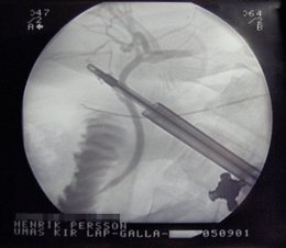
X-Ray during laparascopic cholecystectomy
Find information on thousands of medical conditions and prescription drugs.

Cholecystitis

Cholecystitis is inflammation of the gallbladder. It is commonly due to impaction (sticking) of a gallstone within the neck of the gall bladder, leading to inspissation of bile, bile stasis, and infection by gut organisms. Cholecystitis may be a cause of right upper quadrant pain. The pain may actually manifest in the right flank or scapular region at first. In severe cases, the gall bladder can rupture and form an abscess. In severe cases, it may lead to a life-threatening infection of the liver called cholangitis. In other cases, it may lead to a stable inflammatory state termed chronic cholecystitis. more...

Home
Diseases
A
B
C
Angioedema
C syndrome
Cacophobia
Café au lait spot
Calcinosis cutis
Calculi
Campylobacter
Canavan leukodystrophy
Cancer
Candidiasis
Canga's bead symptom
Canine distemper
Carcinoid syndrome
Carcinoma, squamous cell
Carcinophobia
Cardiac arrest
Cardiofaciocutaneous...
Cardiomyopathy
Cardiophobia
Cardiospasm
Carnitine transporter...
Carnitine-acylcarnitine...
Caroli disease
Carotenemia
Carpal tunnel syndrome
Carpenter syndrome
Cartilage-hair hypoplasia
Castleman's disease
Cat-scratch disease
CATCH 22 syndrome
Causalgia
Cayler syndrome
CCHS
CDG syndrome
CDG syndrome type 1A
Celiac sprue
Cenani Lenz syndactylism
Ceramidase deficiency
Cerebellar ataxia
Cerebellar hypoplasia
Cerebral amyloid angiopathy
Cerebral aneurysm
Cerebral cavernous...
Cerebral gigantism
Cerebral palsy
Cerebral thrombosis
Ceroid lipofuscinois,...
Cervical cancer
Chagas disease
Chalazion
Chancroid
Charcot disease
Charcot-Marie-Tooth disease
CHARGE Association
Chediak-Higashi syndrome
Chemodectoma
Cherubism
Chickenpox
Chikungunya
Childhood disintegrative...
Chionophobia
Chlamydia
Chlamydia trachomatis
Cholangiocarcinoma
Cholecystitis
Cholelithiasis
Cholera
Cholestasis
Cholesterol pneumonia
Chondrocalcinosis
Chondrodystrophy
Chondromalacia
Chondrosarcoma
Chorea (disease)
Chorea acanthocytosis
Choriocarcinoma
Chorioretinitis
Choroid plexus cyst
Christmas disease
Chromhidrosis
Chromophobia
Chromosome 15q, partial...
Chromosome 15q, trisomy
Chromosome 22,...
Chronic fatigue immune...
Chronic fatigue syndrome
Chronic granulomatous...
Chronic lymphocytic leukemia
Chronic myelogenous leukemia
Chronic obstructive...
Chronic renal failure
Churg-Strauss syndrome
Ciguatera fish poisoning
Cinchonism
Citrullinemia
Cleft lip
Cleft palate
Climacophobia
Clinophobia
Cloacal exstrophy
Clubfoot
Cluster headache
Coccidioidomycosis
Cockayne's syndrome
Coffin-Lowry syndrome
Colitis
Color blindness
Colorado tick fever
Combined hyperlipidemia,...
Common cold
Common variable...
Compartment syndrome
Conductive hearing loss
Condyloma
Condyloma acuminatum
Cone dystrophy
Congenital adrenal...
Congenital afibrinogenemia
Congenital diaphragmatic...
Congenital erythropoietic...
Congenital facial diplegia
Congenital hypothyroidism
Congenital ichthyosis
Congenital syphilis
Congenital toxoplasmosis
Congestive heart disease
Conjunctivitis
Conn's syndrome
Constitutional growth delay
Conversion disorder
Coprophobia
Coproporhyria
Cor pulmonale
Cor triatriatum
Cornelia de Lange syndrome
Coronary heart disease
Cortical dysplasia
Corticobasal degeneration
Costello syndrome
Costochondritis
Cowpox
Craniodiaphyseal dysplasia
Craniofacial dysostosis
Craniostenosis
Craniosynostosis
CREST syndrome
Cretinism
Creutzfeldt-Jakob disease
Cri du chat
Cri du chat
Crohn's disease
Croup
Crouzon syndrome
Crouzonodermoskeletal...
Crow-Fukase syndrome
Cryoglobulinemia
Cryophobia
Cryptococcosis
Crystallophobia
Cushing's syndrome
Cutaneous larva migrans
Cutis verticis gyrata
Cyclic neutropenia
Cyclic vomiting syndrome
Cystic fibrosis
Cystinosis
Cystinuria
Cytomegalovirus
Dilated cardiomyopathy
Hypertrophic cardiomyopathy
Restrictive cardiomyopathy
D
E
F
G
H
I
J
K
L
M
N
O
P
Q
R
S
T
U
V
W
X
Y
Z
Medicines

Diagnosis

The classic patient with acute cholecystitis presents with acute right upper quadrant pain, nausea/vomiting, and fever. On physical examination, he or she has a Murphy's sign, which is a diaphragm spasm (due to the intense pain) when the region of the gall bladder is palpated by the examiner.

Laboratory values may be notable for an elevated alkaline phosphatase, possibly an elevated bilirubin (although this could indicate choledocholithiasis), and possibly an elevation of the white blood cell count. The degree of elevation of these laboratory values can be dependent on the degree of inflammation of the gallbladder. Patients with acute cholecystitis are much more likely to manifest abnormal laboratory values, while in chronic cholecystitis the laboratory values are frequently normal.

Radiology

Sonography is a sensitive and specific modality for diagnosis of acute cholecystitis; adjusted sensitivity and specificity for diagnosis of acute cholecystitis are 88% and 80%, respectively. The 2 major diagnostic criteria are cholelithiasis and sonographic Murphy's sign. Minor criteria include gallbladder wall thickening greater than 3mm, pericholecystic fluid, and gallbladder dilatation.

The reported sensitivity and specificity of CT scan findings are in the range of 90-95%. CT is more sensitive than ultrasonography in the depiction of pericholecystic inflammatory response and in localizing pericholecystic abscesses, pericholecystic gas, and calculi outside the lumen of the gallbladder. CT cannot see noncalcified gallbladder calculi, and cannot assess for a Murphy's sign.

Hepatobiliary scintigraphy with technetium-99m bilirubin analogs is also sensitive and accurate for diagnosis of acute cholecystitis, and can differentiate between acute and chronic forms of the disease. It can also assess the ability of the gall bladder to expel bile (gall bladder ejection fraction), and low gall bladder ejection fraction has been linked to chronic cholecystitis. However, since most patients with right upper quadrant pain do not have cholecystitis, primary evaluation is usually accomplished with a modality that can diagnose other causes, as well.

Read more at Wikipedia.org


[List your site here Free!]


MR Cholangiography for Diagnosing Acute Cholecystitis - magnetic resonance
From American Family Physician, 2/15/99 by Barbara Apgar

Ultrasonography has been the screening test of choice for use in the diagnosis of acute cholecystitis. Although ultrasonography is an effective and easily accessible diagnostic modality, it can depict calculi only in the gallbladder and not in the cystic duct. Being aware of the presence of an impacted calculus within the gallbladder neck or cystic duct is important in identifying patients who may develop serious complications of acute cholecystitis. For this reason, other signs, such as gallbladder wall thickness, fluid collection and enlargement of the gallbladder, have been used to aid diagnosis. Magnetic resonance (MR) cholangiography has proved accurate in the diagnosis of bile duct obstruction and choledocholithiasis. Park and associates evaluated the clinical usefulness of MR cholangiography in the diagnosis of acute cholecystitis.

Thirty-five patients with symptoms of acute cholecystitis underwent both ultrasonography and MR cholangiography before cholecystectomy. The images were evaluated, and the results were compared with surgical findings with respect to gallbladder wall thickness and the presence and location of calculi.

Ultrasonography depicted only one of the seven cystic duct calculi (14 percent), but MR cholangiography depicted all of the calculi. There were no false-negative results on cholangiography. One cystic duct calculus suggested on cholangiography was not found at surgery. Fourteen patients had a calculus impacted within the gallbladder neck. Two of the calculi were not demonstrated on ultrasonography. MR cholangiography showed all of the gallbladder neck calculi, and there were no false-negative or false-positive results on imaging of the gallbladder neck.

Eleven patients had floating calculi within the gallbladder lumen. Ultrasonography depicted all of these calculi, but MR cholangiography failed to depict one of them. On a patient-for-patient basis, ultrasonography demonstrated a sensitivity of 62 percent in the diagnosis of cystic duct obstruction, a specificity of 100 percent and an accuracy of 77 percent. MR cholangiography demonstrated a sensitivity of 100 percent, a specificity of 93 percent and an accuracy of 97 percent. In the 29 patients with surgically confirmed thickening of the gallbladder wall, ultrasonography demonstrated a sensitivity of 96 percent, a specificity of 83 percent and an accuracy of 94 percent. MR cholangiography demonstrated a sensitivity of 69 percent, a specificity of 83 percent and an accuracy of 71 percent.

Results of this study demonstrated 100 percent sensitivity for MR cholangiography in the diagnosis of both cystic duct calculi and gallbladder neck calculi. This is much better than the 14 percent and 86 percent sensitivity demonstrated with ultrasonography. The precise location of the calculi was visualized on MR imaging but not on ultrasonography. The Murphy sign, which is useful in the diagnosis of acute cholecystitis on ultrasonography, is not demonstrated on cholangiography. Thus, MR cholangiography should be accompanied by clinical symptoms, laboratory data or ultrasonography to help accurately distinguish acute cholecystitis from chronic cholecystitis.

The authors conclude that MR cholangiography is superior to ultrasonography in the depiction of the causes of cystic duct obstruction, especially cystic duct calculi and calculi in the gallbladder neck. Cost effectiveness of MR cholangiography in this situation remains to be determined. However, when ultrasonography is not diagnostic, MR cholangiography can contribute to management and planning in patients who may develop serious complications of acute cholecystitis.

Park MS, et al. Acute cholecystitis: comparison of MR cholangiography and US. Radiology December 1998; 209:781-5.

COPYRIGHT 1999 American Academy of Family Physicians
COPYRIGHT 2000 Gale Group

Return to Cholecystitis
Home Contact Resources Exchange Links ebay