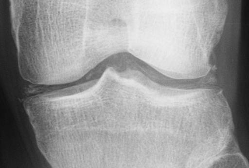
A 65-year-old woman presented with recurrent right knee pain. She had experienced similar persistent and debilitating symptoms 8 months earlier. Evaluation of arthrocentesis on 2 separate occasions was unremarkable. This was followed 5 months later by arthroscopy. Magnetic resonance imaging of the right knee showed lowsignal-intensity lesions compatible with pigmented villonodular synovitis. Biopsy was performed.
Gross examination revealed red to yellow-brown soft tissue fragments, measuring 7 cm in aggregate. Light microscopic examination of formalin-fixed, paraffin-embedded tissue sections stained with hematoxylin-eosin (H&E), revealed hyperplastic synovium lined by a prominent synovial lining with focal edema and fibrosis (Figure 1). The fragments were edematous with focal fibrosis. Immediately beneath the synovial lining were scattered histiocytes and lymphocytes, with some histiocytes containing yellow-brown hemosiderin pigment. This pigment was also noted in the synovial lining. Scattered multinucleated osteoclast-like giant cells were noted. Focally, these giant cells surrounded the pale-staining basophilic crystalline material (Figure 1, c). This material did not polarize in the sections stained with H&E (Figure 2, C). The referring hospital diagnosed the lesion as hyperplastic synovitis suggestive of villonodular tenosynovitis. Sections were stained with the method of Shidham and Shidham.1 Figure 2, A through C show the lesion stained with H&E. Figure 2, D through H show the lesion stained with the Shidham method. Figure 2, parts A, B, E, and F are under ordinary light. Figure 2, parts C, D, G, and H are under polarized light. Figure 2, D and H are under polarized light with slow ray axes perpendicular to each other (direction of the slow ray axis is shown with white arrows with the letter S). The lesion showed rhomboid-shaped and blunt-ended crystals (black arrows) with weak positive birefringence under polarizing microscope in the sections stained with the Shidham method (Figure 2, D and H). The crystals were blue when their long axes were parallel to the slow ray axis and yellow when their long axes were perpendicular to the slow ray axis, using analyzer and polarizing filters (Figure 2, D and H).
What is your diagnosis?
Pathologic Diagnosis: Pseudogout (Chondrocalcinosis Articularis)
Calcium pyrophosphate dehydrate (CPPD) crystals show the diagnostic positive birefringence (Figure 2, D and H) observed in this lesion.2 They are associated with crystal deposition disease, called chondrocalcinosis articularis. This disease manifests with acute or chronic inflammation with joint pain mimicking gout, thus the synonym, pseudogout.3 Sporadic forms of the disease affect people in their sixth to eighth decades of life, rarely before 40 years of age. Pseudogout most often involves the knee and less commonly the wrist or ankle, with sudden onset of severe pain, swelling, and redness with localized heat of involved joint. The arthritis is characteristically monoarticular or oligoarticular.3
In acute pseudogout, synovial fluid usually shows an inflammatory reaction with high white cell counts ranging from 2 to 8 x 10^sup 3^ cells/(mu)L (2000-80000 cells/mm^sup 3^). Eighty percent to 95% of white blood cells are polymorphonuclear cells with intracellular crystals. In patients with chronic or resolving arthritis, the leukocyte count is below 2 x 10^sup 3^ cells/(mu)L (2000 cells/mm^sup 3^), with a predominance of mononuclear cells often with intracellular and extracellular crystals.3 Histologically, pseudogout presents with nodular deposits of CPPD crystals.4-7 Hematoxylineosin-stained sections may not show significant numbers of polarizable crystals owing to the masking of these crystals by hematoxylin in H&E-stained sections (Figure 2, C); however, when the sections are stained with the Shidham method,1,8,9 all the crystals polarize (Figure 2, G) with diagnostic weak positive birefringence (Figure 2, D and H).
Calcium pyrophosphate dihydrate crystals range from 2 to 40 micrometers. They are pleomorphic and are most typically rhomboid shaped, although long or short rods and small squares are also seen. Morphologically, the pseudogout lesions may resemble gout and other conditions, such as tumoral calcinosis.5-7 Pseudogout crystals demonstrate diagnostic weak positive birefringence with polarizing microscopy in sections stained with the Shidham method. The crystals are blue when the long axis of the crystals is parallel to the axis of slow ray and yellow when their long axis is perpendicular to the axis of slow ray. The monosodium urate crystals in gout are present as yellow-brown aggregates of needle-shaped crystals. These crystals demonstrate negative birefringence under polarizing microscopy, with a reverse yellow-blue color pattern as compared to CPPD crystals with reference to the axis of slow ray.1,8,9 In tumoral calcinosis, the calcium hydroxyapatite crystals show amorphous basophilic psammoma body-like calcospherites and do not demonstrate any birefringence in the sections stained with H&E or the Shidham method (V Shidham, unpublished data, July 2000).
An additional diagnostic dilemma may be faced when pseudogout is present in combination with gout and other diseases." In such cases, H&E-stained sections may not detect either CPPD or monosodium urate crystals. If the sections are stained with the Shidham method, both crystals can be detected with their diagnostic birefringence.1,8,9
The authors thank Christopher Chartrand, HT(ASCP), for his expert technological assistance. We also thank Patsy Gill and Sushrut Shidham for their secretarial assistance.
References
1. Shidham V, Shidham G. Staining method to demonstrate urate crystals in formalin-fixed, paraffin-embedded tissue sections. Arch Pathol Lab Med. 2000; 124:774-776.
2. Bullough PG. joint diseases. In: Sternberg SS, Antonioli DA, Mills SE, Carter D, Oberman HA, eds. Diagnostic Surgical Pathology. 3rd ed. Philadelphia, Pa: Lippincott Williams & Wilkins; 1999:230-232.
3. Schumacher HR Jr, Reginato AJ. Atlas of Synovial Fluid Analysis and Crystal Identification. Philadelphia, Pa: Lea & Febiger; 1991:89-132.
4. Bullough PG. Non-neoplastic diseases of bones and joints. In: Silverberg SG, DeLellis RA, Frable WJ, eds. Principles and Practice of Surgical Pathology and Cytopathology. 3rd ed. New York, NY: Churchill Livingstone; 1997:846-847.
5. Ackerman AB, Chongchitnant N, Sanchez J, et al. Histological Diagnosis of Inflammatory Skin Diseases: An Algorithmic Method by Pattern Analysis. 2nd ed. Philadelphia, Pa: Williams & Wilkins; 1997:388.
6. Rosai J. Ackerman's Surgical Pathology. St Louis, Mo: CV Mosby Co; 1996: 1996-1997.
7. Yeldani AA, Kaufman DG, Reddy JK. Cell injury and cellular adaptations. In: Damjanov I, Linder J, eds. Anderson's Pathology. 10th ed. St Louis, Mo: Mosby; 1996:368-369.
8. Shidham VB, Galindo L, Gupta D, Jhala N, Shidham GB. Visualization of urate crystals in paraffin sections [abstract]. Am J Clin Pathol. 1997;107:481.
9. Shidham VB, Galindo LM, Gupta D, Jhala N, Shidham GB. Urate crystals in tissue: a novel staining method for formalin-fixed, paraffin-embedded sections. Lab Med. 1998;29:109-113.
Accepted for publication September 12, 2000.
From the Department of Pathology (Drs Chivukula and V. Shidham) and Nephrology division (Dr G. Shidham), Medical College of Wisconsin, Milwaukee.
Reprints not available from the author.
Copyright College of American Pathologists May 2001
Provided by ProQuest Information and Learning Company. All rights Reserved


