Find information on thousands of medical conditions and prescription drugs.

Dysplasia

Dysplasia (latin for 'bad form') is an abnormality in the appearance of cells indicative of an early step towards transformation into a neoplasia. It is therefore a pre-neoplastic or pre-cancerous change. This abnormal growth is restricted to the epithelial layer, not invading into the deeper tissue. Though dysplasia may regress spontaneously, persistent lesions must be removed, either with surgery, chemical burning, heat burning, burning with laser, or freezing (cryotherapy). more...

Home
Diseases
A
B
C
D
Dandy-Walker syndrome
Darier's disease
Dementophobia
Demyelinating disease
Dendrophobia
Dengue fever
Dental fluorosis
Dentinogenesis imperfecta
Dentophobia
Depersonalization disorder
Dermatitis herpetiformis
Dermatofibroma
Dermatographic urticaria
Dermatomyositis
Dermatophytosis
Desmoplastic small round...
Dextrocardia
Diabetes insipidus
Diabetes mellitus
Diabetes, insulin dependent
Diabetic angiopathy
Diabetic nephropathy
Diabetic neuropathy
Diamond Blackfan disease
Diastrophic dysplasia
Dibasic aminoaciduria 2
Diethylstilbestrol...
DiGeorge syndrome
Dilated cardiomyopathy
Diphallia
Diphtheria
Dipsophobia
Dissociative amnesia
Dissociative fugue
Dissociative identity...
Distemper
Diverticulitis
Diverticulosis
Dk phocomelia syndrome
Doraphobia
Double outlet right...
Downs Syndrome
Dracunculiasis
Duane syndrome
Dubin-Johnson syndrome
Dubowitz syndrome
Duchenne muscular dystrophy
Dupuytren's contracture
Dwarfism
Dysbarism
Dysgerminoma
Dyskeratosis congenita
Dyskinesia
Dysmorphophobia
Dysplasia
Dysplastic nevus syndrome
Dysthymia
Dystonia
E
F
G
H
I
J
K
L
M
N
O
P
Q
R
S
T
U
V
W
X
Y
Z
Medicines

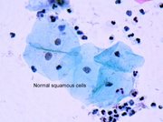
The best know form of dysplasia is the precursor lesions to cervical cancer, called cervical intraepithelial neoplasia (CIN). This lesion is caused by an infection with the human papilloma virus (HPV). Dysplasia of the cervix is almost always unsuspected by the woman. It is usually discovered by a screening test, the pap smear. The purpose of this test is to diagnose the disease early, while it is still in the dysplasia phase and easy to cure.

Dysplasia vs carcinoma in situ vs invasive carcinoma

These terms are related since they represent the three steps of the progression towards cancer:

  • Dysplasia is the earliest form of pre-cancerous lesion recognizable in a biopsy by a pathologist. Dysplasia can be low grade or high grade (see CIS below). The risk of low grade dysplasia transforming into high grade dysplasia and, eventually, cancer is low. Treatment is usually easy.
  • Carcinoma in situ is synonymous with high grade dysplasia in most organs. The risk of transforming into cancer is high. Treatment is still usually easy.
  • Invasive carcinoma, commonly called cancer, is the final step in this sequence. It is a disease who, when left untreated, will invade the host (hence its name) and will probably kill him. It can be often, but not always, be treated successfully.

Metaplasia is a situation where cells have changed from their original mature differentiated type into another mature differentiated cell type as an adaptive response to exposure to chronic irritation, or to a pathogen or carcinogen. It also occurs where one normal cell type changes into another normal cell type as in the cervix where squamous epithelium on the exo-cervix changes to normal columnar epithelium in the endo-cervix. This area is also known as the transformation zone and is the location of many dyplastic lesions thus the sampling of this area during a pap test is critical. Metaplasia is distinct from dysplasia because in a dysplastic cell these changes have become encoded into the genome and so are heritable or passed on to daughter cells during cell replication.

Read more at Wikipedia.org


[List your site here Free!]


Fibrous dysplasia of the frontal sinus
From Ear, Nose & Throat Journal, 1/1/04 by Rafael Rojas

Fibrous dysplasia is a benign idiopathic skeletal disorder that occurs when normal cancellous bone is replaced by abnormal fibrous tissue. The fibrous tissue replaces the spongiosa and fills in the medullary cavity with poorly calcified trabeculae. (1-3)

Fibrous dysplasia represents 2.5% of all bone tumors and 7.5% of all benign bone tumors. (3,4) These tumors usually arise during early childhood or adolescence, and they tend to stabilize after puberty. They occur equally in males and females. Recurrence during adulthood has been noted in approximately 37% of cases. (3,4)

Fibrous dysplasia has two basic clinical forms: monostotic and polyostotic. (1)

The monostotic form, which accounts for approximately 70% of all cases, involves one or two contiguous bones, usually the ribs and the femora. (3) Craniofacial involvement occurs in 10 to 25% of cases; the maxilla and mandibula are most commonly affected. (4)

In the polyostotic form, which accounts for approximately 30% of cases, various areas of the skeleton can be involved; craniofacial involvement occurs in 40 to 50% of cases. (4) Polyostotic fibrous dysplasia, along with endocrine abnormalities and cutaneous hyperpigmentation, is a component of Albright-McCune-Sternberg syndrome, a rare condition that primarily affects females.

Patients with the monostotic form are frequently asymptomatic. They are often diagnosed incidentally during radiographic evaluation for another purpose. Conversely, patients with the polyostotic form have early manifestations, including bone pain and/or bone deformity. These conditions can lead to symptoms of vascular and neurologic compromise, which are easily diagnosed at an early stage.

Craniofacial disease can manifest as headaches and facial distortion (leontiasis ossea). Less severe cases can be characterized by an asymmetric prominence of the face, nasal or sinus obstruction, exophthalmos, and epiphora. Visual and neurologic deficits may occur as a result of cranial nerve involvement. (2,4) Sinus obstruction secondary to fibrous dysplasia may result in infection and the formation of mucoceles.

On computed tomography (CT), the ground-glass appearance of fibrous dysplasia distinguishes it from other lytic lesions (figure 1). (2) Magnetic resonance imaging (MRI) helps evaluate the soft-tissue component, and it can distinguish fibrous dysplasia from other tumors, such as meningiomas (figure 2). Mucocele formation with intracranial or orbital extension is not an uncommon complication of sinus obstruction; this occurs more often in the sphenoid and frontal sinuses and is best assessed by MRI. (2,3)

[FIGURE 1-2 OMITTED]

References

(1.) Bibby K, McFadzean R. Fibrous dysplasia of the orbit. Br J Ophthalmol 1994;78:266-70.

(2.) Daffner RH. Kirks DR, Gehweiler JA, Jr., Heaston DK. Computed tomography of fibrous dysplasia. AM J Roentgenol 1982; 139(5):943-8.

(3.) Jan M, Dweik A, Destrieux C, Djebbari Y. Fronto-orbital sphenoidal fibrous dysplasia. Neurosurgery 1994;34:544-7.

(4.) Som PM, Brandwein M. Tumors and tumor-like conditions. In: Som PM, Curtin HD, eds. Head and Neck Imaging. 4th. ed. St. Louis: Mosby, 2003:331-40.

COPYRIGHT 2004 Medquest Communications, LLC
COPYRIGHT 2004 Gale Group

Return to Dysplasia
Home Contact Resources Exchange Links ebay