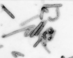
Marburg virus
Find information on thousands of medical conditions and prescription drugs.

Marburg fever

The Marburg virus is the causative agent of Marburg hemorrhagic fever. Both the disease and virus are related to Ebola and originate in the same part of Africa (Uganda and Eastern Congo). The zoonosis is of unknown origin, but some scientists believe it may be hosted by bats. more...

Home
Diseases
A
B
C
D
E
F
G
H
I
J
K
L
M
Mac Ardle disease
Macroglobulinemia
Macular degeneration
Mad cow disease
Maghazaji syndrome
Mal de debarquement
Malaria
Malignant hyperthermia
Mallory-Weiss syndrome
Malouf syndrome
Mannosidosis
Marburg fever
Marfan syndrome
MASA syndrome
Mast cell disease
Mastigophobia
Mastocytosis
Mastoiditis
MAT deficiency
Maturity onset diabetes...
McArdle disease
McCune-Albright syndrome
Measles
Mediterranean fever
Megaloblastic anemia
MELAS
Meleda Disease
Melioidosis
Melkersson-Rosenthal...
Melophobia
Meniere's disease
Meningioma
Meningitis
Mental retardation
Mercury (element)
Mesothelioma
Metabolic acidosis
Metabolic disorder
Metachondromatosis
Methylmalonic acidemia
Microcephaly
Microphobia
Microphthalmia
Microscopic polyangiitis
Microsporidiosis
Microtia, meatal atresia...
Migraine
Miller-Dieker syndrome
Mitochondrial Diseases
Mitochondrial...
Mitral valve prolapse
Mobius syndrome
MODY syndrome
Moebius syndrome
Molluscum contagiosum
MOMO syndrome
Mondini Dysplasia
Mondor's disease
Monoclonal gammopathy of...
Morquio syndrome
Motor neuron disease
Motorphobia
Moyamoya disease
MPO deficiency
MR
Mucopolysaccharidosis
Mucopolysaccharidosis...
Mullerian agenesis
Multiple chemical...
Multiple endocrine...
Multiple hereditary...
Multiple myeloma
Multiple organ failure
Multiple sclerosis
Multiple system atrophy
Mumps
Muscular dystrophy
Myalgic encephalomyelitis
Myasthenia gravis
Mycetoma
Mycophobia
Mycosis fungoides
Myelitis
Myelodysplasia
Myelodysplastic syndromes
Myelofibrosis
Myeloperoxidase deficiency
Myoadenylate deaminase...
Myocarditis
Myoclonus
Myoglobinuria
Myopathy
Myopia
Myositis
Myositis ossificans
Myxedema
Myxozoa
N
O
P
Q
R
S
T
U
V
W
X
Y
Z
Medicines

The disease is spread through bodily fluids, including blood, excrement, saliva, and vomit. There is no cure or vaccine for this deadly and infectious virus. Victims suffer a high fever, diarrhea, vomiting, and severe bleeding from bodily orifices and usually die within a week. Fatality rates range from 25 to 100 %.

In the spring of 2005, the virus attracted widespread press attention for an outbreak in Angola. Beginning in October 2004 and continuing into 2005, the outbreak, which is now thought to be under control, was the world's worst epidemic of any kind of hemorrhagic fever.

The Marburg virus

The viral structure is typical of filoviruses, with long threadlike particles which have a consistent diameter but vary greatly in length from an average of 800 nanometres up to 14,000 nm, with peak infectious activity at about 790 nm. Virions (viral particles) contain seven known structural proteins. While nearly identical to Ebola virus in structure, Marburg virus is antigenically distinct from Ebola virus — in other words, it triggers different antibodies in infected organisms. It was the first filovirus to be identified.

Infection details

Because many of the signs and symptoms of Marburg hemorrhagic fever are similar to those of other infectious diseases, such as malaria or typhoid, diagnosis of the disease can be difficult, especially if only a single case is involved.

The disease is characterised by the sudden onset of fever, headache, and muscle pain after an incubation period of 3-9 days. Within a week, a maculopapular rash develops, followed by vomiting, chest and abdominal pain, and diarrhea. The disease can then become increasingly damaging, causing jaundice, delirium, organ failure, and extensive hemorrhage. Patients generally die from hypovolemic shock as fluid leaks out of the blood vessels, causing blood pressure to drop.

Recovery from the disease is prolonged and can be marked by orchitis, recurrent hepatitis, transverse myelitis or uveitis, or inflammation of the spinal cord, eyes, or parotid gland. Depending upon health care and hospitalization support, the disease can have very high fatality rates, with estimates ranging from 25 % up to 100 %.

Infection is believed to be spread by close contact with body fluids of those infected, and the virus is unlikely to spread through casual contact. Patients are most contagious during the acute phase of the illness when fluids such as vomit and blood are present. Unsafe burial practices such as embracing, kissing or ritual bathing of the corpse present another infection vector.

According to a report in the New York Times, the virus moves very quickly. "On Day 3 of the infection, fewer than 200 viruses are in a drop of blood. By Day 8, there are five million."

Read more at Wikipedia.org


[List your site here Free!]


Ebola and Marburg Viruses: a View of Infection Using Electron Microscopy
From Emerging Infectious Diseases, 8/1/04 by Rollin. Pierre

Elena I. Ryabchikova and Barbara B.S. Price

Batelle Press, Columbus, Ohio ISBN: 1-57477.131-0 Pages: 211, including index Price: US $56.00

More than 25 years have passed since the discovery of a filovirus, Marburg virus, which caused an epidemic of hemorrhagic fever among laboratory workers in Marburg, Germany, in 1967. The persons affected had contact with the blood or tissues of monkeys or with other infected persons. Marburg virus has reappeared only three times since its discovery, with the largest and most recent outbreak occurring in 1999 in Durba, Democratic Republic of the Congo. Ebola virus, another filovirus, was first described in 1976 during two hemorrhagic fever epidemics in Zaire and Sudan. Since then, Ebola virus has caused large hospital outbreaks of hemorrhagic fever in Kikwit, Zaire, in 1995, and Gulu, Uganda, in 2000. Ebola virus has also been implicated in small chains of transmission among persons with direct contact with intermediary hosts, mostly nonhuman primates in the central African countries of Gabon and Republic of the Congo.

The reservoirs for both viruses are still unknown, and the rarity of outbreaks and the remote location of human outbreaks make it difficult, if not impossible, to study the pathogenesis of the human disease. Thus, animal models have been the best, and often only, approach available for studying the progression of disease caused by Marburg and Ebola viruses. Dr. Ryabchikova, the principal author of this book, and her laboratory group have studied the pathogenesis of filoviruses for several decades by using animal models and electron microscopy, a unique approach that has made her one of the few filovirus experts in the world. This book is a compilation not only of her work but of all the information available on Marburg and Ebola viruses.

The first three chapters of the book provide a general review of filovirus history, laboratory methods (with an emphasis on electron microscopy), viral structure, morphology, and replication. Chapters 4 and 5 provide more specific details on infection of the target cells (macrophages and reticulo-endothelial system) in different organs and during the course of filoviral infection. In chapter 6, the authors deal with the "hemorrhagic" side of Ebola and Marburg virus infection. Not all patients infected with these viruses bleed, and when bleeding disorders do occur, no correlated infection of endothelial and hematopoietic cells occurs. Dr. Ryabchikova has found that changes in the microcirculation system, such as the appearance of hemorrhages, clotting, and fibrin deposits, vary by virus and by animal species. Chapter 7, which describes pathologic changes in the organs during the course of filoviral infection, could have been combined with chapter 5. Likewise, the last chapter, which covers immunopathology, appears more like a discussion of the previous chapters.

Much of the data have already been published in the Russian or Western literature. However, this book provides one source for all information available on Marburg and Ebola viruses and has a great advantage over other sources. The number and the quality of the illustrations are impressive, and a comprehensive index is provided. The book will prove useful to clinicians and researchers interested in understanding the pathogenesis of hemorrhagic fevers, and it will provide researchers working with other viruses a lesson in the benefits of using electron microscopy technology.

Pierre Rollin, Centers for Disease Control and Prevention, Atlanta, Georgia, USA

Address for correspondence: Pierre Rollin, Centers for Disease Control and Prevention, 1600 Clifton Road, Mailstop G14, Atlanta, GA, USA; fax: 404-639-1118; email: PRollin@ cdc.gov

COPYRIGHT 2004 U.S. National Center for Infectious Diseases
COPYRIGHT 2004 Gale Group

Return to Marburg fever
Home Contact Resources Exchange Links ebay