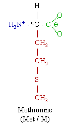
Chemical structure of MethionineChemical structure of methionineimage:Met_biosynthesis.gifimage:Met_pathway.gif
Find information on thousands of medical conditions and prescription drugs.

Methionine

Methionine (Met, M. C5H11NO2S) is an essential nonpolar amino acid, and a lipotropic. more...

Home
Diseases
Medicines
A
B
C
D
E
F
G
H
I
J
K
L
M
Macrodantin
Maprotiline
Marcaine
Marezine
Marijuana
Marinol
Marplan
Matulane
Maxair
Maxalt
Maxolon
MDMA
Measurin
Mebendazole
Mebendazole
Meclofenoxate
Medrol
Mefenamic acid
Mefloquine
Melagatran
Melarsoprol
Meloxicam
Melphalan
Memantine
Metadate
Metamfetamine
Metamizole sodium
Metandienone
Metaxalone
Metenolone
Metformin
Methadone
Methamphetamine
Methaqualone
Metharbital
Methcathinone
Methenamine
Methionine
Methocarbamol
Methohexital
Methotrexate
Methotrexate
Methoxsalen
Methylcellulose
Methyldopa
Methylergometrine
Methylin
Methylphenidate
Methylphenobarbital
Methylprednisolone
Methyltestosterone
Methysergide
Metiamide
Metoclopramide
Metohexal
Metoprolol
Metrogel
Metronidazole
Metyrapone
Mobic
Moclobemide
Modafinil
Modicon
Monopril
Montelukast
Motrin
Moxidectin
Moxifloxacin
Moxonidine
MS Contin
Mucinex
Mucomyst
Mupirocin
Mupirocin
Muse
Mycitracin
Mycostatin
Myfortic
Mykacet
Mykinac
Myleran
Mylotarg
Mysoline
Phentermine
N
O
P
Q
R
S
T
U
V
W
X
Y
Z

Methionine and cysteine are the only sulfur-containing proteinogenic amino acids. The methionine derivative S-adenosyl methionine (SAM) serves as a methyl donor. Methionine plays a role in cysteine, carnitine and taurine synthesis by the transsulfuration pathway, lecithin production, the synthesis of phosphatidylcholine and other phospholipids. Improper conversion of methionine can lead to atherosclerosis. Methionine is a chelating agent.

Methionine is one of only two amino acids encoded by a single codon (AUG) in the standard genetic code (tryptophan, encoded by UGG, is the other). The codon AUG is also significant, in that it carries the "Start" message for a ribosome to begin protein translation from mRNA. As a consequence, methionine is incorporated into the N-terminal position of all proteins in eukaryotes and archaea during translation, although it is usually removed by post-translational modification. Methionine can also occur at other positions in the protein.

Foods containing methionine include fruits, vegetables, and legumes. High levels of methionine can be found in spinach, green peas, corn, navel and mandarin oranges, peanuts, pistachios, macadamia nuts, kidney beans, black turtle beans, tofu, and tempeh.

Biosynthesis

Since methionine is an essential amino acid, it cannot be synthesized in humans. However, in plants and microorganisms, methionine is synthesized from aspartic acid and cysteine. First, aspartic acid is converted to β-aspartyl-semialdehyde, an important intermediate in the biosynthesis of methionine, lysine, and, threonine. Of homoserine by homoserine acyltransferase, puts a good leaving group on homoserine allowing it to react with cysteine to produce cystathionine. Enzymatic cleavage of cystathionine yilds homocysteine, which can then be methylated by folates to give methionine. Both cystathionine-γ-synthase and cystathionine-β-lyase require Pyridoxyl-5'-phosphate as a cofactor, while homocysteine methyltransferase requires Vitamin B12 as a cofactor.

Methionine is converted to S-adenosylmethionine (SAM) by (1) methionine adenosyltransferase. SAM serves as a methyl-donor in many (2) methyltransferase reactions and is conveted to S-adenosylhomocysteine (SAH). (3) adenosylhomocysteinase converts SAH to homocysteine.

Read more at Wikipedia.org


[List your site here Free!]


Relationship between S-adenosyl-methionine, S-adenosylhomocysteine, asymmetric dimethylarginine, and endothelial function in healthy human subjects during
From Alternative Medicine Review, 6/1/05 by S. Doshi

Doshi S, McDowell I, Goodfellow J, et al. Metabolism 2005;54:351-360.

Experimental hyperhomocysteinemia after an oral methionine or homocysteine load is associated with impaired nitric oxide-dependent vasodilatation in healthy human beings. However, it remains unproven that this effect is mediated by elevations in plasma homocysteine. There is evidence that an increase in plasma homocysteine may increase the formation of asymmetric dimethylarginine (ADMA), an inhibitor of nitric oxide synthase. The methyl groups within ADMA are derived from the conversion of S-adenosylmethionine to S-adenosylhomocysteine intermediates in the methionine/homocysteine pathway. No previous study has assessed the role of methylation status, its impact on ADMA formation, and their association with endothelial function in healthy human beings. In a randomized, placebo-controlled, crossover study, 10 healthy subjects (mean age, 29.1 +/- 3.9 years) were administered an oral dose of methionine (0.1 g/kg), L-homocysteine (0.01 g/kg), N-acetylcysteine (NAC) (0.1 g/kg), or placebo. Endothelial function as assessed by flow-mediated dilatation (FMD) of the brachial artery was impaired after both the methionine and homocysteine load compared with placebo at 4 hours (36 +/- 15, 67 +/- 23 vs 219 +/- 26 microm, respectively, P < .001). N-acetylcysteine had no effect on flow-mediated dilatation. Plasma total homocysteine was significantly elevated at 4 hours after methionine (23.1 +/- 6.2) and homocysteine (41.5 +/- 8.9) loading, but significantly reduced after NAC 2.4 +/- 0.6 vs 7.1 +/- 2.1 micromol/L in the placebo (P < .001). Plasma S-adenosylmethionine/S-adenosylhomocysteine ratio was significantly (P < .001) increased at 4 hours after methionine (10.9 +/- 0.7) compared with homocysteine (5.4 +/- 0.4), NAC (5.0 +/- 0.3), and placebo (6.0 +/0.5). Plasma ADMA concentrations were not altered by any intervention. Our results suggest that endothelial dysfunction due to methionine or homocysteine loading is not associated with an increase in plasma ADMA or a disruption in methylation status.

COPYRIGHT 2005 Thorne Research Inc.
COPYRIGHT 2005 Gale Group

Return to Methionine
Home Contact Resources Exchange Links ebay