Norwegian scabies may be difficult to diagnose clinically, and this may have serious consequences. The presence of peripheral and tissue eosinophilia in a patient with a rash should prompt exhaustive searches for the underlying cause (box), and clinicians should remember that findings from skin biopsy specimens can be unreliable in obtaining an accurate diagnosis. We describe the consequences of misdiagnosis in an elderly patient with Norwegian scabies whose rash was atypical and was considered to be the result of a drug reaction.
Causes of various types of eosinophilia
* Infecfious--helminths
* Respiratory--eosinophilic pneumonitis, asthma
* Gastrointestinal--inflammatory bowel disease
* Allergic--asthma, eczema
* Systemic--vasculitis
* Iatrogenic--drugs
* Malignant--lymphoma, colonic carcinoma
Case report
A 77 year old man was admitted to hospital from a nursing home in August 1997. He had had a widespread, red, itchy, and scaly rash for 4 weeks. The man had been generally unwell and anorexic, but showed no specific features of systemic disease. He had previously been diagnosed as having senile dementia, congestive cardiac failure, and Barrett's oesophagus. The patient had been taking bumetanide (1 mg daily), captopril (12.5 mg twice daily), fluoxetine (20 mg daily), and lanzoprazole (20 mg daily) long term (for at least 6 months). In addition, his general practitioner had prescribed amoxycillin and an antihistamine for the rash.
A presumptive diagnosis of an adverse drug reaction was made, and captopril or bumetanide were suspected. These two drugs and the lanzoprazole were stopped, and the man was treated with a moderately potent topical steroid containing an antibiotic. His urea and electrolyte values were normal, but his albumin concentration was slightly low at 29 g/1 (normal range 36-52 g/l). Thyroid function and liver enzyme activities were normal; the patient's chest radiograph showed slight pulmonary plethora, and an electrocardiogram showed no acute abnormal features. The patient had a raised total white cell count and a noticeably high peripheral eosinophilia (4.1x[10.sup.9]/1 (normal range 0.0-0.4x[10.sup.9]/l)). Microscopic investigation of a stool sample showed no abnormal findings. Examination of a skin biopsy specimen showed spongiosis, exocytosis, and a moderate perivascular inflammatory skin infiltrate with numerous eosinophils extending to the mid-dermis (fig 1). Given the clinical setting and the details presented to the histopathologist, these biopsy findings were thought to be consistent with an adverse drug reaction.
[Figure 1 ILLUSTRATION OMITTED]
The patient improved gradually and was discharged to the nursing home 5 weeks later. At this time his skin condition had largely resolved, though the peripheral eosinophilia persisted. He was readmitted to hospital 4 days later, profoundly confused as a result of staphylococcal septicaemia. The septicaemia had developed because his rash had become infected. This resolved with antibiotic treatment, and the man was discharged after 3 days. However, he still had a dry, crusted rash.
The skin condition worsened gradually, and the man was readmitted to hospital 2 months later. At this time the rash was lichenified and hyperkeratotic, with scale formation on the hands and feet. Skin scrapings were taken and these showed plentiful scabetic mites. The patient was treated twice with benzyl benzoate to the body and permethrin to the head and neck. The rash resolved quickly and for the first time since his initial admission to hospital the patient's eosinophil count fell to within the normal range.
There had been an outbreak of scabies among the ward staff and several fellow residents in the nursing home after the patient's initial discharge from hospital in August 1997, some 5 months before the final diagnosis. The skin biopsy specimen was re-examined in the light of the further clinical information and it was confirmed that the appearances could be consistent with scabies infection, although no mites were seen.
Discussion
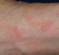
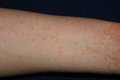
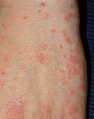
Crusted (Norwegian) scabies is a highly contagious variant of ordinary scabies that is known to affect elderly patients,[1] especially those with cognitive deficiency? It can present atypically and mimic a range of other dermatoses, including eczema and psoriasis; widespread hyperkeratotic crusted lesions are often seen (fig 2). Norwegian scabies affects people who are immunosuppressed and it is relatively common in HIV positive patients, in whom it may be diagnosed mistakenly as "pruritus of AIDS."[3] Misdiagnosis is associated with two serious consequences--the eruption may become infected secondarily (box), particularly with Staphylococcus aureus, and someone with an undiagnosed condition may infect numerous contacts. Both of these problems occurred with our patient. Indeed, in a similar case, a patient had been misdiagnosed as having drug induced erythroderma for 2 years before the diagnosis of Norwegian scabies was made.[4] Again, retrospective epidemiological investigation showed that this patient had infected several contacts.[4]Scabies infection can have serious consequences if accurate diagnosis is delayed or missed altogether, especially in high risk or debilitated patients in whom secondary bacterial infection can give rise to life threatening complications.[5]
Clinical features of scabies
* Rash--polymorphic: papulovesicular/excematous/ pustular
* Itch--especially nocturnal and after hot shower
* Secondary bacterial infection--usually Staphylococcus aureus; nephritogenic streptococci can cause glomerulonephritis
Skin biopsy specimens from patients with scabies usually show a dermal infiltrate of lymphocytes and histiocytes, with or without neutrophils and eosinophils, and denser inflammation in the nodular lesions. Skin biopsy specimens from people with Norwegian scabies almost always show plentiful mites and eggs, especially in the thickened stratum corneum within areas of hyperkeratotic skin. The biopsy specimen from our patient did not show hyperkeratosis, eggs, or mites, but it was taken before the typical features of Norwegian scabies rash, which he displayed on his latter hospital admission, had developed. In the clinical context in which the biopsy specimen was taken, the histological features were thought to be consistent with a drug rash. However, these features were not specific and in retrospect it was felt that the appearances would also have been in keeping with those of scabies.
Obtaining skin scrapings is useful in diagnosing suspected scabies and should be the initial investigation.[6] The technique is best performed by scraping the skin with a round bladed scalpel in order to remove flakes of tissue, having first put a drop of oil on the affected area. Diagnostic yield is improved if scraping is carried out over scabetic papules and burrows, which are commonly seen in the web spaces of the fingers. The scrapings are placed directly on a microscope slide and viewed under magnification (x20) for the presence of characteristic Sarcoptes scabiei mite's eggs or faecal pellets. If the slides have to be sent to the laboratory, the coverslip should be sealed with nail varnish.
Benzoyl benzoate (25%) used to be the most commonly used scabicidal agent in United Kingdom, but because it causes irritation in children malathion or permthrin are more often used in younger patients.[6] These drugs also have the advantage that a single application is usually enough. In severe scabies infestation, several application are often needed, and the scalp should also be treated.[6] More recently, crusted scabies in HIV positive and negative patients has been treated successfully with a single oral dose of the antihelminthic agent ivermectin.[7] However, this drug has not yet been licensed in the United Kingdom and there have been doubts about its safety in elderly patients.[8]
Our patient almost certainly had scabies at the time he was misdiagnosed as having a drug rash. It is possible that the rash was caused by something else initially and that he became infected secondarily with scabies, but the fact that the eosinophilia, which had been present since his first presentation, settled after specific treatment for scabies suggests that he had scabies all along. From the evolution of the rash and the appearance of the skin biopsy specimen, we suspect that this case of scabies developed into the Norwegian variant we have described here.
[1] Hopper AH, Salisbury J, Jegadeva AN, Scott B, Bennett GC. Epidemic Norwegian scabies in a geriatric unit. Age Ageing 1990; 19:125-7.
[2] Kolar KA, Rapini RP. Crusted (Norwegian) scabies. Am Fam Physician 1991;44:1317-21.
[3] Sclesinger I, Oelrich DM, Tyring SK. Crusted (Norwegian) scabies in patients with AIDS: the range of clinical presentations. South Med J 1994;87:352-6.
[4] Shelley WB, Shelley ED, Burmeister V. Staphylococcus aureus colonisation of burrows in erythrodermic Norwegian scabies. A case study of iatrogenic contagion. J Am Acad Dermatol 1988; 19:673-8.
[5] Bonomo RA, Jacobs M, Jacobs G, Graham R, Salata RA. Norwegian scabies and a toxic shock syndrome toxin 1-producing strain of Staphylococcus aureus endocarditis in a patient with trisomy 21. Clin Infect Dis 1998;27:645-6.
[6] Burgess IF. Diagnosing and treating scabies. Practitioner 1996;240:739-43.
[7] Meinking TL, Taplin D, Hermida JL, Pardo R, Kerdel FA. The treatment of scabies with ivermectin. N Engl J Med 1995;333:26-30.
[8] Barkwell R, Shields S. Deaths associated with ivermectin treatment of scabies. Lancet 1997;349:1144-5.
(Accepted 31 March 1999)
Department of Clinical Pharmacology and Therapeutics, University of Liverpool, Liverpool L69 3BX
D S Almond senior registrar
School of Pharmacy, John Moores University, Liverpool L3 3AF
C J Green research pharmacist
Royal Liverpool and Broad Green University Hospitals NHS Trust, Liverpool L7 8XP
D M Geurin consultant, department of pathology
S Evans consultant, department of determatology
Correspondence to: D S Almond solly@liv.ac.uk
BMJ 2000;320:35-6
COPYRIGHT 2000 British Medical Association
COPYRIGHT 2000 Gale Group


 Home
Home A
A Sabinas brittle hair...