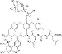
Methicillin-resistant Staphylococcus aureus (MRSA) with reduced vancomycin susceptibility vancomycin-intermediate S. aureus (VISA) has been reported from many countries. Whether resistance is evolving regularly in different genetic backgrounds or in a single clone with a genetic predisposition, as early results suggest, is unclear. We have studied 101 MRSA with reduced vancomycin susceptibility from nine countries by multilocus sequence typing (MLST), characterization of SCCmec (staphylococcal chromosomal cassette mec), and agr (accessory gene regulator). We found nine genotypes by MLST, with isolates within all five major hospital MRSA lineages. Most isolates (88/101) belonged to two of the earliest MRSA clones that have global prevalence. Our results show that reduced susceptibility to vancomycin has emerged in many successful epidemic lineages with no clear clonal disposition. Increasing antimicrobial resistance in genetically distinct pandemic clones may lead to MESA infections that will become increasingly difficult to treat.
**********
Methicillin-resistant Staphylococcus aureus (MRSA) is a major problem around the world, causing hospital-acquired infections and, more recently, infections in the community (1,2). The glycopeptides, particularly vancomycin, have been the mainstays of therapy for MRSA, and the emergence of resistance to these agents is of great concern.
The first S. aureus with reduced vancomycin susceptibility (vancomycin MIC [greater than or equal to] 8 [micro]g/mL) was isolated in 1997 (3,4), and similar isolates have since been discovered in several countries. These vancomycin-intermediate S. aureus (VISA) isolates are relatively rare; a recent review found 21 VISA described in the literature (5). However, strains of S. aureus have been described that are vancomycin-susceptible by conventional testing but have a subpopulation of resistant cells. These heterogeneous VISA (hVISA) are more common; reports from around the world indicate that 0.5%-20% of MESA are heteroresistant (5). The clinical importance of hVISA is debatable, but evidence shows that they are precursors of VISA, and they have been implicated in treatment failure in deep-seated infections (6,7).
A study of early VISA strains that used multilocus sequence typing (MLST) and analysis of the SCCmec region suggested that they were all descended from the New York/Japanese (8,9) pandemic MESA clone (10); the first high-level vancomycin-resistant isolates that have acquired the vanA gene cassette from enterococci are also members of this clone (F. Tenover, pers. comm.). Researchers have suggested that isolates of the New York/Japanese pandemic MESA clone may be predisposed to become vancomycin resistant, perhaps because of loss-of-function mutations in the agr (accessory gene regulator) gene (11). We analyzed the genetic backgrounds of a geographically diverse sample of VISA and hVISA to investigate the evolutionary history of such strains.
Materials and Methods
We collected 101 isolates of MESA with reported heterogeneous or homogeneous resistance to vancomycin (MIC [greater than or equal to] 8 mg/L) from China (n = 1), France (31), Japan (2), Norway (14), Poland (13), Sweden (1), United Kingdom (28), and the United States (11). Antimicrobial susceptibility tests were performed by the agar dilution method of the National Committee for Clinical Laboratory Standards. Isolates were described as VISA if they fulfilled the three criteria adopted by the Centers for Disease Control and Prevention, that is, broth microdilution vancomycin MIC of 8 to 16 mg/L, MIC [greater than or equal to] 6 mg/L on E-test, and growth on brain-heart infusion agar containing 6 mg/L vancomycin (12). Isolates with heterogeneous resistance to vancomycin were confirmed by using population analysis profiling followed by measuring the area under the curve (PAP-AUC), as described previously (13). The prototypic hVISA strain MU3 was used as a standard, and isolates with an AUC [greater than or equal to] 0.9 compared to MU3 were described as hVISA.
MLST was performed as described previously (10). The seven housekeeping gene sequences were compared to known alleles in the MLST database (available from http://www.mlst.net), and the resulting allelic profiles (which define sequence types, STs) were used to interrogate the databases for matches within records of the 988 isolates held there. The MLST databases contain molecular and epidemiologic data on S. aureus isolates from carriage and disease, including examples of all major MRSA clones (10). Data from this study were added to the S. aureus MLST database, and the entire dataset was analyzed by using the BURST algorithm to assign isolates to clonal complexes (CCs), which are lineages containing genetically related isolates (sharing 100% genetic identity at [greater than or equal to] 5/7 loci used). Polymerase chain reaction (PCR) analysis of the ccr (chromosomal cassette recombinase) and mec (methicillin resistance) regions was performed to discriminate the four main SCCmec types (I-IV) on the basis of combinations of the two regions. Conventional PCR was used to detect SCCmec I-III by using the primers described in Ito et al. (14) and SCCmec IV by using those described by Daum et al. (15). These results were confirmed using the multiplex method of Oliveira et al. (16). Detection of agr subgroups I-IV was performed by PCR of the region surrounding agrD, which codes for an autoinducing peptide, according to the method of Peacock et al. (17).
Results and Discussion
The results are shown in the Table. PAP-AUC values for the isolates varied from 0.9 to 3.01 and 91/101 isolates were designated hVISA on the basis of a PAP-AUC value >0.9. Nine isolates were designated as VISA.
From the genotyping results, strains were divided into clonal complexes, which can be subdivided according to sequence type (ST) and SCCmec differences. The clonal complexes CC5, CC8, CC22, CC30, and CC45 represent the five pandemic MRSA lineages that have been previously described (10). Our results show that hVISA has arisen in all five of these pandemic clones and that VISA has so far developed in CC5 and CC8. The three most common MRSA clones present in the United Kingdom (EMRSA-3, EMRSA-15, EMRSA-16) (18) are included within these lineages, and reduced vancomycin susceptibility has been identified in all of these clones. All lineages displayed resistance to multiple antimicrobial classes, and only the new oxazolidinone linezolid was active against all strains.
Only agr subgroups (alleles) I and II were found in isolates in this study with 7/9 VISA and 57/92 hVISA having agr I. Within the 14 clones in this study, the proportion of isolates with particular agr alleles was variable. The presence of both agr I and agr II among VISA/hVISA, even in genetically similar isolates, suggests that the genes for the agr system are horizontally transferred. Sakoulas et al. reported an association of agr II with the development of vancomycin resistance (11). Our results show that VISA/hVISA also emerged in strains with agr I.
Molecular analyses of VISA isolates to date have focused on isolates from the United States and Japan, and results have indicated that all strains belong to the New York/Japanese MRSA clone. In our study, we found that hVISA isolates have emerged from every lineage that has produced pandemic MRSA clones, and VISA isolates have emerged in two of five lineages, in all likelihood from hVISA precursor isolates.
Increasing drug resistance in clones that are multidrug resistant and adapted to spread and cause serious disease can do much damage in the modern hospital environment. We have shown that reduced vancomycin susceptibility has emerged in genetically and phenotypically diverse MRSA clones throughout the world. This finding suggests that vancomycin resistance has the potential to become a widespread problem in MRSA strains already resistant to multiple antimicrobial agents.
References
(1.) Chambers HF. The changing epidemiology of Staphylococcus aureus? Emerg Infect Dis 2001;7:178-82.
(2.) Herold BC, Immergluck LC, Maranan MC, Lauderdale DS, Gaskin RE, Boyle-Vavra S, et al. Community-acquired methicillin-resistant Staphylococcus aureus in children with no identified predisposing risk. JAMA 1998;279:593-8.
(3.) Centers for Disease Control and Prevention. Update: Staphylococcus aureus with reduced susceptibility to vancomycin--United States, 1997. MMWR Morb Mortal Wkly Rep 1997;46:813-5.
(4.) Hiramatsu K, Hanaki H, Ino T, Yabuta K, Oguri T, Tenover FC. Methicillin-resistant Staphylococcus aureus clinical strain with reduced vancomycin susceptibility. J Antimicrob Chemother 1997;40:135-6.
(5.) Walsh TR, How RA. The prevalence and mechanisms of vancomycin resistance in Staphylococcus aureus. Annu Rev Microbiol 2002;56:657-75.
(6.) Ariza J, Pujol M, Cabo J, Pena C, Fernandez N, Linares J, et al. Vancomycin in surgical infections due to methicillin-resistant Stapylococcus aureus with heterogeneous resistance to vancomycin. Lancet 1999;353:1587-8.
(7.) Moore MR, Perdreau-Remington F, Chambers HF. Vancomycin treatment failure associated with heterogeneous vancomycin-intermediate Staphylococcus aureus in a patient with endocarditis and in the rabbit model of endocarditis. Antimicrob Agents Chemother 2003;47: 1262-6.
(8.) Ito T, Katayama Y, Hiramatsu, K. Cloning and nucleotide sequence determination of the entire mec DNA of pre-methicillin-resistant Staphylococcus aureus N315. Antimicrob Agents Chemother 1999;43:1449-58.
(9.) Oliveira DC, Tomasz A, de Lencastre H. The evolution of pandemic clones of methicillin-resistant Staphylococcus aureus: identification of two ancestral genetic backgrounds and the associated mec elements. Microb Drug Res 2001;7:349-61.
(10.) Enright MC, Robinson DA, Randle G, Fell EJ, Grundmann H, Spratt BG. The evolutionary history of methicillin-resistant Staphylococcus aureus (MRSA). Proc Natl Acad Sci U S A 2002;99:7687-92.
(11.) Sakoulas G, Eliopoulos GM, Moellering RC Jr, Wennersten C, Venkataraman L, Novick RP, et al. Accessory gene regulator (agr) locus in geographically diverse Staphylococcus aureus isolates with reduced susceptibility to vancomycin. Antimicrob Agents Chemother 2002;46:1492-502.
(12) Tenover FC, Biddle JW, Lancaster MV. Increasing resistance to vancomycin and other glycopeptides in Staphylococcus aureus. Emerg Infect Dis 2001 ;7:327-32.
(13.) Wootton M, Howe RA, Hillman R, Walsh TR, Bennett PM, MacGowan AP. A modified population analysis profile (PAP) method to detect hetero-resistance to vancomycin in Staphylococcus aureus in a UK hospital. J Antimicrob Chemother 2001;47:399-403.
(14.) Ito T, Katayama Y, Asada K, Mori N, Tsutsumimoto K, Tiensasitorn C, et al. Structural comparison of three types of staphylococcal cassette chromosome mec integrated in the chromosome in methicillin-resistant Staphylococcus aureus. Antimicrob Agents Chemother 2001;45:1323-36.
(15.) Daum RS, Ito T, Hiramatsu K, Hussain F, Mongkolrattanothai K, Jamklang M, et al. A novel methicillin-resistance cassette in community-acquired methicillin-resistant Staphylococcus aureus isolates of diverse genetic backgrounds. J Infect Dis 2002;186:1344-7.
(16.) Oliveira DC, de Lencastre H. Multiplex PCR strategy for rapid identification of structural types and variants of the mec element in methicillin-resistant Staphylococcus aureus. Antimicrob Agents Chemother 2002;46:2155-61.
(17.) Peacock S J, Moore CE, Justice A, Kantzanou M, Story L, Mackie K, et al. Virulent combinations of adhesin and toxin genes in natural populations ofStaphylococcus aureus. Infect Immun 2002;70:4987-96.
(18.) Epidemic methicillin resistant Staphylococcus aureus. Commun Dis Rep CDR Wkly 1997;7:191.
This work was funded by the Wellcome Trust. M.C.E. is a Royal Society University Research Fellow.
Dr. Howe is a consultant microbiologist at Southmead Hospital, Bristol, and Clinical Lecturer at Bristol University. His research interests include many areas of clinical microbiology, particularly the mechanisms of antimicrobial resistance in bacterial pathogens.
Address for correspondence: Mark C. Enright, Department of Biology and Biochemistry, University of Bath, Bath, BA2 7AY, UK; fax: +441225-386779; email: m.c.enright@bath.ac.uk
Robin A. Howe, * Alastair Monk, ([dagger]) Mandy Wootton, * Timothy R. Walsh, ([double dagger]) and Mark C. Enright ([dagger])
* Southmead Hospital, Bristol, United Kingdom; ([dagger]) University of Bath, Bath, United Kingdom; and ([double dagger]) University of Bristol, Bristol, United Kingdom
COPYRIGHT 2004 U.S. National Center for Infectious Diseases
COPYRIGHT 2004 Gale Group


