Two patients with von Recklinghausen disease (neurofibromatosis type 1) were admitted to the hospital because of progressive heart failure. Both patients had prominent pulmonary hypertension revealed on cardiac catheterization. A lung perfusion scan did not show any gross defect. There were no underlying causes of pulmonary hypertension in either patient, such as chronic lung disease, congenital or acquired heart disease, deep vein thrombosis, or systemic hypercoagulable states. There may be an unrecognized association between von Recklinghausen disease and pulmonary hypertension.
(CHEST 2001; 119:1606-1608)
Key words: epoprostenol; neurofibromatosis; plasma brain natriuretic peptide; pulmonary hypertension; vasculopathy; von Recklinghausen disease
Abbreviations: BNP = brain natriuretic peptide; MAA = macroaggregated albumin; NYHA = New York Heart Association
Von Recklinghausen disease (neurofibromatosis type 1) is an autosomal dominant disorder that can be recognized by characteristic skin lesions, cafe-au-lait spots, and cutaneous neurofibromas.[1] The disease is occasionally complicated with systemic vasculopathy, such as renovascular hypertension, myocardial infarction, cerebral infarction, ischemic bowel disease, or rupture of an arterial aneurysm.[2,3] Only one case of pulmonary artery involvement has been reported up to now.[4]
We report here two cases of von Recklinghausen disease complicated with prominent pulmonary hypertension. Vasculopathy of von Recklinghausen disease might underlie pulmonary hypertension in the two patients.
CASE REPORT
Case 1
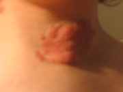
The first patient was a 19-year-old Japanese woman who had received a diagnosis of von Recklinghausen disease from her skin lesions at 5 years of age. She had progressive dyspnea, palpitation, and general fatigue on effort at the age of 15 years. Fourteen months later, she had an episode of chest pain and syncope when she rode a bicycle. Her first admission to our hospital was at the age of 16 years. She was in New York Heart Association (NYHA) functional class 2. Systemic multiple cafe-au-lait spots and a right lumbar cutaneous neurofibroma were noticed. She had no history of sleep apnea, appetite suppressant use, or solvent inhalation. An accentuated pulmonic component of the second heart sound was audible, but lung sounds were normal. Her chest radiograph showed slightly dilated central pulmonary arteries. ECG revealed right ventricular hypertrophy, QT prolongation, and inverted T waves in leads aVL and [V.sub.2] through [V.sub.5]. An echocardiogram showed right atrial and right ventricular enlargement. Mild indentation of the interventricular septum toward the left ventricle was found. Left-sided valvular structures and left ventricular size and function were normal. A Doppler echocardiographic study represented moderate tricuspid regurgitant flow, and right ventricular systolic pressure was estimated at 72 mm Hg. Antiphospholipid syndrome and collagen diseases were excluded by serology findings. Arterial blood gas analysis obtained in room air showed [PCO.sub.2] of 39 mm Hg and [PO.sub.2] of 67 mm Hg at rest. Respiratory function tests showed [FEV.sub.1] of 2.8 L (84% predicted), vital capacity of 3.4 L (116% predicted), and diffusion capacity of the lung for carbon monoxide of 21.9 mL/min/mm Hg (79% predicted). Lung CT showed no evidence of pulmonary disease. Proximal pulmonary arteries were dilated. Obstruction or obliteration of the arterial lumen was not detected. A [sup.99m]Tc-macroaggregated albumin (MAA) lung perfusion scan showed small perfusion defects (Fig 1). Cardiac catheterization demonstrated a pulmonary artery pressure of 84/31 mm Hg, cardiac index of 2.1 L/min/[m.sup.2], pulmonary capillary wedge pressure of 12 mm Hg, total pulmonary resistance of 1,210 dyne [multiplied by] s [multiplied by] [cm.sup.-5], and systemic arterial pressure of 102/65 mm Hg (Table 1). Primary pulmonary hypertension was diagnosed, complicated by von Recklinghausen disease. The patient was treated with nifedipine and warfarin because nifedipine reduced total pulmonary resistance 24% without significant systemic BP lowering at the acute challenge test. Her symptoms gradually worsened after discharge, and she was admitted this time at 19 years of age. Arterial blood gas status on room air showed a [PCO.sub.2] of 27 mm Hg and [PO.sub.2] of 74 mm Hg. An echocardiogram revealed that the right ventricle and the right atrium were markedly enlarged and that the left ventricle was extremely compressed by the right ventricle. A Doppler echocardiographic study showed severe tricuspid regurgitation and pulmonary regurgitation, and right ventricular systolic pressure was estimated at 113 mm Hg. At cardiac catheterization, pulmonary artery pressure was 117/54 mm Hg, cardiac index was 1.4 L/min/[m.sup.2], pulmonary capillary wedge pressure was 11 mm Hg, and total pulmonary resistance was 2,833 dyne [multiplied by] s [multiplied by] [cm.sup.-5]. Epoprostenol was administered IV at a dosage of 8 ng/kg/min for 3 months; her dyspnea, palpitation, and chest discomfort on effort improved gradually. Plasma brain natriuretic peptide (BNP) level, which was commonly used as an indicator of congestive heart failure, fell from 1,017 pg/mL before the epoprostenol therapy to 91 pg/mL 3 months later.[5] Functional capacity improved from the NYHA functional class 3 to class 2. She received oral beraprost instead of IV epoprostenol, and then she returned home.
[ILLUSTRATION OMITTED]
Case 2
The second case was a 70-year-old Japanese woman with von Recklinghausen disease. The patient had an appendectomy at the age of 15 years, had been treated for hypertension since 56 years of age, and had borderline diabetes mellitus. She had no history of appetite suppressant use, sleep apnea, or solvent inhalation. There was no episode of acute pulmonary embolism or deep vein thrombosis. She has been admitted three times for heart failure in the past. On her first admission (at 66 years of age), chest radiography disclosed moderate cardiomegaly with a cardiothoracic ratio of 63%, dilated central pulmonary arteries, and pulmonary congestion. Clinical examination did not reveal deep venous thrombosis. Functional capacity was NYHA functional class 3. Arterial blood gas analysis on room air showed severe hypoxemia ([PCO.sub.2], 34 mm Hg; [PO.sub.2], 48 mm Hg). An ECG showed incomplete right bundle-branch block and right ventricular hypertrophy. Serum transaminase levels were normal. Blood coagulation abnormalities were not found. Serum HIV antibody was not detected. An echocardiogram disclosed right ventricular hypertrophy and moderately compressed left ventricle by the enlarged right ventricle. Left ventricular wall motion and left-sided valvular structures were normal. Doppler echocardiographic study showed mild tricuspid regurgitation. Cardiac catheterization demonstrated pulmonary artery pressure of 70/22 mm Hg (mean, 38 mm Hg), pulmonary capillary wedge pressure of 8 mm Hg, cardiac index of 2.3 L/min/[m.sup.2], total pulmonary resistance of 956 dyne [multiplied by] s [multiplied by] [cm.sup.-5], and systemic arterial pressure of 132/78 mm Hg (Table 1). Congenital heart diseases were not disclosed. A lung perfusion scan with [sup.99m]Tc-MAA showed slightly decreased accumulation at peripheral areas of the right segment 1, 2, and left segment 1 + 2 (Fig 2). The extent of perfusion defects was not related to the severity of pulmonary hypertension. She had no respiratory diseases and no deep vein thrombosis, and primary pulmonary hypertension was diagnosed. She received warfarin, isosorbide dinitrate, diuretics, and home oxygen therapy. However, her symptoms progressively worsened, and she was admitted to the hospital a second time, 23 months later. Although treatment with oral beraprost was started, symptoms were not improved. At the time of her third hospital admission (at 69 years of age), her heart failure became refractory and her functional capacity declined to NYHA functional class 4. Echocardiography showed severely pressed left ventricle by the right ventricle, and giant right-sided chambers. Doppler echocardiographic study showed moderate tricuspid regurgitation and mild pulmonary regurgitation. Cardiac catheterization demonstrated pulmonary artery pressure of 96/30 mm Hg (mean, 55 mm Hg), cardiac index 1.4 L/min, and systemic BP of 132/78 mm Hg. Acute challenge test of epoprostenol showed a fall in pulmonary artery mean pressure (22%) and increase in cardiac index (33%) without significant systemic BP reduction during cardiac catheterization. She received continuous epoprostenol infusion therapy at a dosage of 5 to 8 ng/kg/min. Her symptoms improved 3 months later. BNP that was considered as a marker of congestive heart failure decreased markedly (1,507 to 253 pg/mL).[5]
[ILLUSTRATION OMITTED]
DISCUSSION
The prognosis is generally good in most patients with von Recklinghausen disease, but the course is occasionally complicated with systemic vasculopathy that results in renovascular hypertension, myocardial infarction, cerebral infarction, ischemic bowel disease, or rupture of an arterial aneurysm.[2,3] Such vascular complications are major determinants of the morbidity and mortality in patients with von Recklinghausen disease.[3] Samuels et al[4] described a case complicated with pulmonary hypertension, which is rare in this disease. In that case, multiple pulmonary thromboemboli were initially suspected by lung perfusion scintigraphy and pulmonary arteriography. However, pulmonary endarterectomy disclosed primary vasculopathy with no evidence of thromboembolism. There was another report that described complications of pulmonary hypertension secondary to pulmonary fibrosis in von Recklinghausen disease.[6]
We describe here two cases of von Recklinghausen disease complicated by pulmonary hypertension. Respiratory organ diseases, hypercoagulable states, or other diseases that might have caused pulmonary hypertension were not disclosed. There were no gross or segmental defects on [sup.99m]Tc-MAA lung perfusion scans. Thus, primary pulmonary hypertension was diagnosed. Because both primary pulmonary hypertension and von Recklinghausen disease are rare disorders, the possibility of chance complications is low, though it cannot be completely excluded. There may be a causal relationship between these disorders.
Pulmonary endothelial dysfunction is recently believed to play a role in the pathophysiology of primary pulmonary hypertension, and it leads to vasoconstriction and in situ thrombosis.[7-9] The vasoconstriction will lead to medial hypertrophy and/or plexiform lesions. In systemic vasculopathy of von Recklinghausen disease, intimal hypertrophy with smooth muscle cell proliferation, aneurysmal dilatation of small arteries, and plexiform lesions have been demonstrated.[3] Therefore, vasculopathy of von Recklinghausen disease might underlie the pathophysiology of pulmonary hypertension in our two patients, although we could not confirm it by histology.
REFERENCES
[1] Riccardi VM. von Recklinghausen neurofibromatosis. N Engl J Med 1981; 305:1617-1627
[2] Lehrnbecher T, Gassel AM, Rauh V, et al. Neurofibromatosis presenting as a severe systemic vasculopathy. Eur J Pediatr 1994; 153:107-109
[3] Lie JT. Vasculopathies of neurofibromatosis type 1 (von Recklinghausen disease). Cardiovasc Patho] 1998; 7:97-108
[4] Samuels N, Berkman N, Milgalter E, et al. Pulmonary hypertension secondary to neurofibromatosis: intimal fibrosis versus thromboembolism. Thorax 1999; 54:858-859
[5] Tsutamoto T, Wada A, Maeda K, et al. Attenuation of compensation of endogenous cardiac natriuretic peptide system in chronic heart failure. Circulation 1997; 96:509-516
[6] Porterfield JK, Pyeritz RE, Traill TA. Pulmonary hypertension and interstitial fibrosis in von Recklinghausen neurofibromatosis. Am J Med Genet 1986; 25:531-535
[7] Rich S. Clinical insights into the pathogenesis of primary pulmonary hypertension. Chest 1998; 114:237S-241S
[8] Pietra GG, Edwards WD, Kay JM, et al. Histopathology of primary pulmonary hypertension: a qualitative and quantitative study of pulmonary blood vessels from 58 patients in the National Heart, Lung, and Blood Institute, Primary Pulmonary Hypertension Registry. Circulation 1989; 80: 1198-1206
[9] Yuan JX, Aldinger AM, Juhaszova M, et al. Dysfunctional voltage-gated [K.sup.+] channels in pulmonary artery smooth muscle cells of patients with primary pulmonary hypertension. Circulation 1998; 98:1400-1406
(*) From the First Department of Internal Medicine (Drs. Aoki, Kodama, Mezaki, and Aizawa), Niigata University School of Medicine, Niigata City; and Department of Cardiology (Drs. Ogawa, Sato, and Okabe), Tachikawa General Hospital, Nagaoka City, Japan.
Manuscript received February 29, 2000; revision accepted November 21, 2000.
Correspondence to: Yoshinori Aoki, MD, First Department of Internal Medicine, Niigata University School of Medicine, 1-757, Asahimachi-dori, Niigata City 951-8510, Japan
COPYRIGHT 2001 American College of Chest Physicians
COPYRIGHT 2001 Gale Group


