Definition
Wolff-Parkinson-White syndrome is an abnormality in the electrical functioning of the heart which may cause rapid heart rates. The abnormality affects the electrical signal between the atria and ventricles.
Description
Blood is circulated through the heart and body by a muscular pump and valve system involving the atria and ventricles. The right atrium receives oxygen-lacking blood returning to the heart from the body. The blood is passed from the right atrium into the right ventricle, which contracts and sends blood out to the pulmonary artery. The pulmonary artery sends the blood into the lungs, where carbon dioxide is removed, and fresh oxygen is added. The left atrium receives blood with oxygen from the lungs and passes this arterial blood to the left ventricle, where it is emptied into the aorta, the main artery of the heart.
These functions are directed by electrical signals within the heart. In patients afflicted with Wolff-Parkinson-White syndrome, an abnormal pathway exists that causes additional electrical signals to pass between the atria and ventricles, possibly causing rapid heart rate.
Causes & symptoms
Congenital heart disease may contribute to this and other arrhythmias. Ebstein's anomaly, a congenital heart defect that involves displacement of the tricuspid valve, located on the right side of the heart, is one known cause of Wolff-Parkinson-White syndrome. This anomaly allows blood to flow via the small hole to the other side of the heart. Often, there is no known cause for Wolff-Parkinson-White syndrome. Many people with the syndrome have no symptoms. On the other hand, some people experience temporary rapid heartbeat due to certain drugs, smoking, and anxiety.
Diagnosis
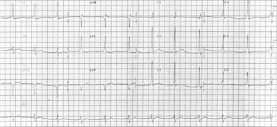
Electrocardiography (ECG) is used to diagnose Wolff-Parkinson-White syndrome, and other cardiac arrhythmias. A trained physician, normally a cardiologist, can recognize patterns of electrical conduction. With this syndrome, the extra pathway will show a pattern different from those of normal conduction. If no irregular patterns show on the ECG, the patient may be sent home with a 24-hour heart monitor, called a Holter monitor, which will help detect intermittent occurrences. Other studies, such as the cardiac electrophysiologic study (EPS), may be ordered to pinpoint the location of the accessory pathway, and to determine a course of treatment.
Treatment
Various drugs may be used to treat Wolff-Parkinson-White syndrome, as well as other cardiac arrhythmias. The purpose of these drugs is to slow the electrical signals and excitation of heart muscles. As some of these drugs may have side effects, including the rare production of new or more frequent arrhythmias, the patient should be carefully observed. Ablative therapies may be accomplished with radiofrequency or cardiac catheters to cut through the tissue which is causing the abnormal electrical signals.
At one time, only open heart surgery was used, but the procedure can be done now with local anesthesia in a special cardiac laboratory. In some cases, surgery may still be recommended to treat Wolff-Parkinson-White syndrome.Young people with this syndrome may be treated more successfully with surgery, rather than enduring a lifetime of drug treatments, or the possible threat of sudden cardiac death.
Alternative treatment
A provider may teach patients methods to help control heart rate. Relaxation techniques, acupuncture, botanical medicine, and homeopathy can all be helpful supportive therapies.
Prognosis
Most patients with this syndrome can lead normal lives, even with episodes of tachycardia. In many cases, the syndrome is secondary to the underlying congenital heart defect. However, Wolff-Parkinson-White syndrome can cause sudden cardiac arrest in certain instances.
Prevention
If the syndrome is not due to congenital heart disease, the patient may try avoiding behaviors which lead to arrhythmia, such as elimination of caffeine, alcohol, cocaine, and smoking.
Key Terms
- Ablative
- Used to describe a procedure involving removal of a tissue or body part, or destruction of its funtion.
- Arrhythmia
- Irregular heart beat.
- Electrocardiograph (ECG)
- A test of a patient's heartbeat that involves placing leads, or detectors, on the patient's chest to record electrical impulses in the heart. The test will produce a strip, or picture record of the heart's electrical function.
- Tachycardia
- Rapid heart rate, defined as more than 100 beats per minute.
Further Reading
For Your Information
Books
- Conn, Howard F., Robert J. Clohecy, and Rex B. Conn. Current Diagnosis. Philadelphia: W.B. Saunders, 1997.
Organizations
- American Heart Association. 7320 Greenville Ave. Dallas, TX 75231. (214) 373-6300. http://www.amhrt.org.
- National Institutes of Health. National Heart, Lung and Blood Institute. Building 31, Room 4A21, Bethesda, MD 20892. (301) 496-4236. http://www.nhlbi.nih.gov.
Gale Encyclopedia of Medicine. Gale Research, 1999.


