Abstract
We report the case of a patient with neurofibromatosis type 1 who had both aplasia of an internal carotid artery (ICA) and a vagal neurofibroma. To our knowledge, this is the first report in the literature of the simultaneous presence of these two rare disorders in a single patient. We believe that this is also the first report of an absence of an ICA in a patient with neurofibromatosis type 1. The patient was a 19-year-old woman who complained of a slowly growing neck mass. The mass occupied the right parapharyngeal space and upper cervical region. The patient had no other masses on physical examination, but widespread cafe au lait spots were evident. This led us to suspect the presence of a vagal neurofibroma. The tumor was removed, and pathology confirmed the diagnosis. No intracranial aneurysms were detected on cerebral angiography.
Introduction
Agenesis of the internal carotid artery (ICA) is a rare congenital disorder that was first described by Tode in 1787. [1] Only about 100 cases have been reported to date. The first radiologic demonstration of this anomaly was made by Verbiest during his angiographic study in 1954. [1]
Neurofibroma of the vagus is also rare; when it does occur, it can involve any part of the nerve throughout its cervical course. [2,3] Vagal neurofibromas are usually slow-growing, asymptomatic tumors. They can appear sporadically or in association with neurofibromatosis (von Recklinghausen's disease). [3]
In this article, we describe the case of a patient with neurofibromatosis type 1 who had an absence of an ICA (on the left) and a vagal neurofibroma (on the right). To our knowledge, this is the first report in the literature of these two disorders occurring in the same patient and the first report of an ICA aplasia in a patient with neurofibromatosis type 1.
Case report
A right-handed, 19-year-old woman complained of a mass on the right side of her neck that had been present for 12 months. She was admitted to the hospital. She had no personal history of hoarseness, seizures, or benign neural neoplasms and no family history of neurofibromatosis.
On otolaryngologic examination, her ears, nose, laryngeal structures, and both vocal folds were normal. On oropharyngeal examination, a minimal medial displacement of the right palatine tonsil and soft palate was evident. Findings on transnasal endoscopic examination of the nasopharynx were normal.
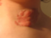
Visual inspection revealed that the woman had bilateral axillary freckling and multiple cafe au lait spots larger than 2 cm on her back, abdomen, and both pretibial regions. On palpation, a firm, semimobile, painless, well-circumscribed, nonpulsatile, 4-cm mass was detected medial to the right mandibular angle and upper cervical region deep to the sternocleidomastoid muscle. No other neck mass, lymphadenopathy, or cutaneous neurofibroma was palpable. Results of a neurologic examination were normal except for a minor motor deficit in the right upper extremity.
Computed tomography (CT) of the neck revealed a well-circumscribed, heterogeneous, hypodense, 4.5-cm mass between the jugular foramen and the root of the neck on the right at the level of the oropharynx (figure 1). No cervical lymph nodes were apparent.
During our review of the CT image, we noted that the bony canal for the ICA on the left was hypoplastic. Therefore, we ordered selective angiography of the carotid and vertebral arteries to document the status of both ICAs as well as to ascertain the vascular nature of the mass. Angiography revealed that the left ICA was absent (figure 2). Left anterior and middle cerebral arterial flow was maintained through the anterior communicating artery. The vertebral arteries and the right internal and external carotid arteries were patent and normal. No intracranial aneurysm was present. As for the mass itself, it appeared to displace the right ICA medially, but it was not vascular.
Cranial CT demonstrated a left cerebral hemiatrophy and a secondary enlargement of the left lateral ventricle. Cranial CT confirmed the earlier neck CT finding that the left bony canal for the ICA was hypoplastic (figure 3).
The presence of the cafe au lait spots and the CT appearance of the mass led us to suspect that the patient had a neural tumor, and surgical resection was planned. (CT is mandatory during the preoperative assessment of any parapharyngeal mass. [2]) The mass was surgically removed via a transcervical approach. Intraoperatively, it became evident that the grayish-pink mass had originated in the vagus nerve and had displaced the right ICA medially and the internal jugular vein posteriorly. The mass was bluntly dissected from the major vessels of the neck, but it was not possible to dissect it from the vagus nerve. Therefore, the vagus nerve was severed at the level of the clavicle. The lateral traction of the mandible enabled us to dissect the mass in the parapharyngeal space. The entire mass was excised. No major bleeding occurred. Histopathology confirmed that the mass was a neurofibroma (figure 4).
Postoperatively, the woman experienced tachycardia (which responded well to beta blocker therapy), right vocal fold paralysis, hoarseness, and a minimal degree of dysphagia. Aspiration was not a major problem. She was discharged from the hospital on postoperative day 10 without any complications.
Discussion
ICA anomalies. Congenital ICA anomalies can be classified as agenic, aplastic, or hypoplastic. The absence of the ICA is the result of either agenesis or aplasia. The term a genesis is used when both the ICA and its bony canal are absent; aplasia refers to a situation where the ICA is absent but there is some evidence of a bony canal.[4] Our patient had ICA aplasia, because angiography showed that the ICA was absent while CT showed that a small portion of the bony canal was present. Intracranial aneurysms have been reported in 25% of patients with ICA agenesis. [5] No intracranial aneurysm was detected in our patient.
Patients with ICA anomalies can be asymptomatic. In such cases, the diagnosis can be delayed until a neurologic manifestation appears. When symptoms are present, they are caused by cerebrovascular insufficiency, compression of the brain by vessels that were dilated to compensate for the absence of the ICA, or the presence of an aneurysm. [4] Our patient's chief complaint was the presence of the cervical mass; her complaint had no relation to her ICA aplasia. Aside from the presence of the mass, she was asymptomatic, except for the minimal motor deficit in her right upper extremity.
Vagal neurofibromas. Vagal neurofibromas have been reported sporadically as well as in patients with neurofibromatosis. [3] Our patient had neurofibromatosis type 1. These rare tumors exhibit no predilection toward either sex. [6] Patients are usually asymptomatic, although dysphagia or hoarseness might be evident secondary to Xth cranial nerve paralysis. [2,3,6]
Agenesis of an ICA has been reported in patients with neurofibromatosis type 2, [7] but until now there has been no report of its occurrence in either a patient with neurofibromatosis type 1 or a patient with a vagal neurofibroma.
Because our patient had no ICA on the left, it was important that we preserve the ICA on the right during surgical resection of the mass on the right, and this we were able to accomplish.
The hemicerebral atrophy in our patient was presumed to be related to vascular insufficiency caused by the aplasia of the left ICA. Another case of hemicerebral atrophy with an absence of an ICA has been reported in the literature. [8]
Neurofibromas have been reported to undergo a malignant transformation in approximately 8% of patients with neurofibromatosis. [9] Irradiation has been reported to cause neurofibromas to change into a neurofibrosarcomas in patients with neurofibromatosis. [10]
Surgical resection is the treatment of choice for a vagal neurofibroma. Because it is a benign tumor, the surgeon should attempt to preserve the nerve. This might not be possible when the tumor absorbs nerve axons as it grows. [9]
Acknowledgment
The authors gratefully acknowledge the kind assistance provided by Dr. Murat Ozcan during the preparation of the article.
References
(1.) Worthington C, Olivier A, Melanson D. Internal carotid artery agenesis: Correlation by conventional and digital subtraction angiography, and by computed tomography. Surg Neurol 1984;22:295-300.
(2.) Peetermans JF, Van de Heyning PH, Parizel PM, et al. Neurofibroma of the vagus nerve in the head and neck: A case report. Head Neck 1991;13:56-61.
(3.) Green JD, Jr., Olsen KD, DeSanto LW, Scheithauer BW. Neoplasms of the vagus nerve. Laryngoscope 1988;98:648-54.
(4.) Claros P, Bandos R, Gilea I, et al. Major congenital anomalies of the internal carotid artery: Agenesis, aplasia and hypoplasia. Int J Pediatr Otorhinolaryngol 1999;49:69-76.
(5.) Cali RL, Berg R, Rama K. Bilateral internal carotid artery agenesis: A case study and review of the literature. Surgery 1993;113:227-33.
(6.) Galli J, Almadori G, Paludetti G, et al. Plexiform neurofibroma of the cervical portion of the vagus nerve. J Laryngol Otol 1992;106:643-8.
(7.) Chen MC, Liu HM, Huang KM. Agenesis of internal carotid artery associated with neurofibromatosis type II. AJNR Am J Neuroradiol 1994;15:1184-6.
(8.) Afifi AK, Godersky JC, Menezes A, et al. Cerebral hemiatrophy, hypoplasia of internal carotid artery, and intracranial aneurysm. A rare association occuring in an infant. Arch Neurol 1987;44:232-5.
(9.) Moore G, Yarington CT, Jr., Mangham CA, Jr. Vagal body tumors: Diagnosis and treatment. Laryngoscope 1986;96:533-6.
(10.) Ducatman BS, Scheithauer BW. Postirradiation neurofibrosarcoma. Cancer 1983;51:1028-33.
COPYRIGHT 2001 Medquest Communications, Inc.
COPYRIGHT 2001 Gale Group


