Catabolism is increased after burn injury, characterized by significant loss of body protein and bone. Long-term consequences include growth arrest in children, low bone mineral density, and increased incidence of fracture.
Vitamin D is an important nutrient in children due to its role in bone formation during growth and development. It facilitates intestinal absorption of calcium and is required for calcium and phosphorus homeostasis. The prevalence of vitamin D insufficiency, its etiology and associated sequelae among acutely injured burn patients is unknown. Therefore, a recent study examined vitamin D and endocrine status, as well as the effect of anabolic agents, in acutely injured pediatric patients who had sustained burns in excess of 25% total body surface area (TBSA).
Sixty-nine patients with a mean TBSA burn of 50.6% and full thickness injury of 41.3% were studied. Subjects ranged in age from 0.6 yr to 18 yr. Morning blood samples were obtained weekly for serum 25-hydroxyvitamin D (D25), 1,25-dihydroxyvitamin D (D1,25), albumin, cortisol, triodothyronine (T3), tetraiodothyronine (T4), thyroid stimulating hormone (TSH) and parathormone (PTH). Exogenous vitamin D intake was not controlled in this study. Patients consumed vitamin Dad lib from an oral diet, plus they typically received twice the RDA of vitamin D per liter of tube feeding. Hypovitaminosis D was assessed by dividing patients into three diagnostic categories according to their serum vitamin D concentrations: those with adequate vitamin D25 status (>37.4 umol/L) or D 1,25 (>38.8 pmol/L), those with low vitamin D (20-37.3 nmol/L or D 1,25 (20.8-38.8 pmol/L)and those with very low serum vitamin D (<20 umol/L for D25, < 20.8 pmol/L for D1,25).
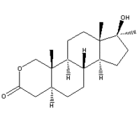
A total of 280 morning blood samples of D25 and D 1,25 demonstrated that 45% and 26.2% were low and 8.9% and 11% were very low, respectively. At least one low D25 or D1,25 level occurred in 62.3% of all children. Very low serum vitamin D levels were noted in 23.2% of all patients. There was an increased incidence of hyperparathyroidism in patients with very low serum D25. Vitamin D25 and D 1,25 levels were lower in subjects with larger burns or inhalation injury, as well as those treated with thyroxine or oxandrolone. Serum albumin, cortisol, T4, and TSH were not correlated with concentration of vitamin D.
Demonstration of a high incidence of low serum vitamin D indicates vitamin D status may be significantly compromised in burned children. One reason why this may be true is that serum albumin is the major binding protein of D1,25. Serum albumin levels decrease and remain low for long periods after thermal injury. However, there was no relationship found between albumin and D 1,25 in this study. It is also conceivable that the bum wound and scarring and also a lack of sunlight exposure may limit cutaneous vitamin D biosynthesis. Further studies should be conducted to determine whether larger doses of vitamin D, earlier initiation of treatment or perhaps a different form of vitamin D may be required to obtain improvements in outcome measures postburn.
Michele M. Gottschlich, Theresa Mayes, Jane Khoury, and Glenn D. Warden, Hypovitaminosis D in acutely injured pediatric bum patients, JADA 104(6): 931-934 (June 2004) [Address correspondence to: Michele M. Gottschlich, PhD, RD, Shriners Hospitals for Children, Nutrition Services, 3229 Burnet Ave, Cincinnati, OH 45229. E-mail: mgottschlich@shrinenet.org]
COPYRIGHT 2004 Frost & Sullivan
COPYRIGHT 2004 Gale Group


