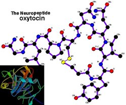
Oxytocin structure. Inset shows oxytocin bound to neurophysin
Find information on thousands of medical conditions and prescription drugs.

Oxytocin

Oxytocin is a mammalian hormone that in women is released mainly after stimulation of the nipples or distention of the vagina and that facilitates birth and breastfeeding. It is also released during orgasm in both sexes. In the brain, it acts as a neurotransmitter and is involved in bonding and the formation of trust between people. more...

Home
Diseases
Medicines
A
B
C
D
E
F
G
H
I
J
K
L
M
N
O
Methoxsalen
OCL
Octabenzone
Octanoic acid
Octopamine
Octreotide
Ofloxacin
Ofloxacin
Olanzapine
Omeprazole
Oncovin
Ondansetron
Opium
Oratane
Oretic
Orinase
Orlistat
Ornidazole
Ornithine
Orotic acid
Orphenadrine
Ortho Evra
Oruvail
Ovcon
Ovral
Ovrette
Oxaliplatin
Oxamniquine
Oxandrin
Oxandrolone
Oxaprozin
Oxazepam
Oxcarbazepine
Oxetine
Oxibendazole
Oxiracetam
Oxprenolol
Oxybenzone
Oxybuprocaine
Oxycodone
Oxycodone
Oxymetazoline
Oxymetholone
Oxymorphone
Oxytetracycline
Oxytocin
P
Q
R
S
T
U
V
W
X
Y
Z

Synthetic oxytocin is sold as medication under the trade names Pitocin and Syntocinon and also as generic Oxytocin.

Synthesis, storage and release

Oxytocin is made in magnocellular neurosecretory cells in the supraoptic nucleus and paraventricular nucleus of the hypothalamus and is released into the blood from the posterior lobe of the pituitary gland. Oxytocin is also made by some neurons in the paraventricular nucleus that project to other parts of the brain and to the spinal cord.

In the pituitary gland, oxytocin is packaged in large, dense-core vesicles, where it is bound to neurophysin as shown in the inset of the figure; neurophysin is a large peptide fragment of the giant precursor protein molecule from which oxytocin is derived by enzymatic cleavage.

Secretion is regulated by the electrical activity of the oxytocin cells in the hypothalamus. These cells generate action potentials that propagate down axons to the neurosecretory nerve endings in the pituitary; the endings contain large numbers of oxytocin-containing vesicles, which are released by exocytosis when the terminals are depolarised.

Structure and relation to vasopressin

Oxytocin is a peptide of nine amino acids (a nonapeptide). The sequence is cysteine - tyrosine - isoleucine - glutamine - asparagine - cysteine - proline - leucine - glycine (CYIQNCPLG). The cysteine residues form a sulfur bridge.

Oxytocin has a molecular mass of 1007 daltons. One international unit (IU) of oxytocin is the equivalent of about 2 micrograms of pure peptide.

The structure of oxytocin is very similar to that of vasopressin, which is also a nonapeptide with a sulfur bridge. Oxytocin and vasopressin are the only known hormones released by the human posterior pituitary gland to act at a distance. However, oxytocin neurons can make corticotropin-releasing hormone (CRH) and vasopressin neurons dynorphin, for example, that act locally. The magnocellular neurons that make oxytocin are adjacent to magnocellular neurons that make vasopressin, and are similar in many respects.

Oxytocin and vasopressin were discovered, isolated and synthesized by Vincent du Vigneaud in 1953, work for which he received the Nobel Prize in Chemistry in 1955.

The oxytocin receptor is a G-protein-coupled receptor which requires Mg2+ and cholesterol. It belongs to the rhodopsin-type (class I) group of G-protein-coupled receptors.

Actions

Oxytocin has peripheral (hormonal) actions, and also has actions in the brain.

Peripheral (hormonal) actions

The peripheral actions of oxytocin mainly reflect secretion from the pituitary gland. Oxytocin receptors are expressed by the myoepithelial cells of the mammary gland, and in both the myometrium and endometrium of the uterus at the end of pregnancy. In some mammals, oxytocin receptors are also found in the kidney and heart.

Read more at Wikipedia.org


[List your site here Free!]


Use of Oxytocin to Shorten Labor and Avoid C-Sections
From American Family Physician, 2/15/00 by Anne D. Walling

Approximately 25 percent of deliveries in the United States are induced or augmented by oxytocin, but there is little consensus regarding the optimal dosage regimen. Recommendations for the initial dose range from 0.5 to 6.0 mU per minute, with dosage adjustments ranging from 1 to 6 mU per minute. The recommended interval between adjustments also varies from 15 to 60 minutes. Typically, the end points in studies of augmented labor are time to delivery and need for cesarean delivery caused by fetal distress related to hyperstimulation of the uterus. However, these studies are technically challenging because of difficulties associated with blinding caretakers and addressing ethical concerns regarding the safety of mother or fetus. Merrill and Zlatnik compared the impact of high and low doses of oxytocin on duration of labor and need for cesarean delivery in women who required induction or augmentation of labor.

All women beyond 24 weeks' gestation who were admitted to a university obstetric unit for augmented labor and delivery were eligible for the study. The women had to have medical indications for augmented labor and no contraindications to oxytocin therapy or augmentation of delivery. Those who met the study criteria were randomized to receive high- or low-dose oxytocin. The solutions were prepared in the pharmacy, and obstetric staff had no knowledge of the patient's group assignment. Patients in the low-dose group received 5 U of oxytocin per 500 mL of solution, while those in the high-dose group received 15 U of oxytocin per 500 mL of solution. The initial infusion rate in both groups was 9 mL per hour. Patients in the low-dose group received 1.5 mU per minute initially, which was then increased by 1.5 mU per minute every 30 minutes until adequate labor was established. High-dose patients received 4.5 mU per minute, which was then increased by 4.5 mU per minute every 30 minutes. The infusion was discontinued if severe fetal heart rate abnormalities occurred or if more than seven contractions were documented in a 15-minute period. If no progress was documented after eight to 12 hours, the induction was regarded as "failed," and alternative treatments could be used.

A total of 816 patients was included in the analysis: 412 patients in the low-dose group and 404 in the high-dose group. Patient age, race, gestation, parity and indication for induction were similar between groups. Despite randomization, more multiple births were noted in the low-dose group, but these accounted for less than 4 percent of patients. The mean time to achieving full dilation was significantly reduced in the high-dose group (7.8 hours) compared with the low-dose group (9.7 hours). The mean time to delivery was also significantly reduced (8.5 compared with 10.5 hours). Women in the high-dose group also had a lower rate of cesarean delivery (11.3 compared with 15 percent), but this difference was not statistically significant. Oxytocin was reduced or discontinued twice or more in 41 percent of the high-dose group and in 30 percent of the low-dose group. The side effect panel, including placental abruption, infection, postpartum hemorrhage and length of maternal hospital stay, was similar between groups. One woman in each group experienced uterine rupture; both cases were associated with previous cesarean delivery. Neonatal outcomes were also similar between groups, including Apgar score, birth weight, acidosis and length of hospital stay.

The authors conclude that high-dose oxytocin significantly shortens labor and tends to be associated with fewer cesarean deliveries. In addition, the use of a high dose does not appear to increase or intensify the side effects associated with oxytocin.

Merrill DC, Zlatnik FJ. Randomized, double-masked comparison of oxytocin dosage in induction and augmentation of labor. Obstet Gynecol September 1999;94:455-63.

COPYRIGHT 2000 American Academy of Family Physicians
COPYRIGHT 2000 Gale Group

Return to Oxytocin
Home Contact Resources Exchange Links ebay