Cystine is the chief sulfur-containing amino acid of protein molecules, and it is an important source of sulfur in the metabolism. Cystinuria is an uncommon autosomal recessive disorder of metabolism that results in decreased reabsorption of four amino acids (ie, cystine, lysine, ornithine, arginine) from the renal tubule. Only cystine forms calculi. Urinary cystine calculi form because of the low solubility of cystine in urine with a pH less than 7.0. Approximately one in 20,000 individuals is diagnosed with urinary calculi, and 1% to 4% of urinary calculi are cystine calculi.(1)
Patients are diagnosed with cystinuria if they have
* an early onset of clinical calculus disease,
* a family history of recurrent calculus disease, and
* recurrent calculus formation unresponsive to the usual forms of therapy.(2)
The urine of patients diagnosed with cystinuria has characteristic hexagonal crystals, along with concentrations of cystine in excess of 75 mg/g creatinine.(3)
MEDICAL MANAGEMENT
Medical management of these patients is based on three principals:
* decreasing total cystine concentration in urine,
* increasing the solubility of cystine in urine, and
* increasing urinary excretion of cystine.(4)
Therapeutic measures include fluid diuresis, alkalinization of the urine to a pH of 7.5 with sodium bicarbonate or sodium potassium citrate, and use of cystine-binding medications (eg, D-penicillamine, [Alpha]-mercaptopropionylglycine). This regimen is difficult to maintain, however, due to frequent voiding and the medications' side effects (eg, rash, fever, agranulocytosis, iron depletion, proteinuria, nephrotic syndrome). Dietary restrictions also tend to consist of bland, poorly tolerated foods.(5) Methionine (ie, a sulfur-bearing compound) is restricted in patients' diets, but because it is an essential amino acid found in protein, it is difficult for patients to completely eliminate it from their diets. For these reasons, many patients continue to develop urinary calculi throughout their lifetimes.
SURGICAL TREATMENT
Surgical treatment of ureteral cystine calculi includes cystoscopy with basket retrieval of the calculi. If the calculi are large or are impacted in the patient's ureter, conservative treatment includes stenting the ureter to cause dilation so the calculi may pass on their own. Ureterolithotomy (ie, incising the ureter and removing the obstructing calculus) is considered an option only when other medical-surgical treatments have failed. This procedure requires a flank or abdominal incision, and recovery time can be extensive.
An ileal ureter transplant is performed if a patient develops large or recurrent calculi that require continuous treatment. During this procedure, the surgeon removes the affected ureter through a midline incision and replaces it with a portion of the patient's small intestine. Due to the large size of the intestinal lumen, calculi no longer become lodged in the ureter. This procedure is not without complications, however. Urinary reflux occurs in 25% of patients, even when the ileum is rotated 90 degrees to make it isoperistaltic (ie, allow bowel contents to move in the usual direction) and the intestine is tapered when it is implanted. Late developing strictures also have been reported.(6)
FLASH LAMP PULSED DYE LASER TREATMENT
The flash lamp pulsed dye laser was developed in 1967. The pulsed dye laser has a wavelength of 504 nanometers (nary) in duration pulses of approximately 1 millisecond, is green, and is absorbed by the yellow pigment of most urinary calculi without elevating the temperature of the calculi more than 50 [degrees] F (10 [degrees] C).(7) This laser's wavelength is absorbed minimally by body tissue or hemoglobin, and it is safe to use on impacted calculi. The process of laser energy being absorbed by the calculi creates a "plasma," or small cloud of highly excited ions and electrons.(8) This plasma rapidly expands and contracts at the rate of pulsation (ie, 5 to 10 hertz [Hz]), causing an acoustic shock wave from the irrigation used during the procedure.(9) It is this mechanical action that breaks the calculi into passable fragments.
The pulsed dye laser uses a 320-micron (pm) fiber passed through a flexible ureteroscope. The maximum power for the flash lamp pulsed dye laser is 140 millijoules, with a repetition rate of 10 Hz. The pulsed dye laser works extremely well on calcium oxalate calculi. Cystine calculi absorb only minimal amounts of light, which is insufficient to permit the formation of a photoacoustic effect (ie, a mechanical shock wave that is produced by laser light).(10)
HOLMIUM: YTTRIUM-ALUMlNUM-GARNET LASER TREATMENT
The holmium:yttrium-aluminum-garnet (YAG) laser is a solid-state crystal of YAG doped (ie, a process by which elements are suspended within a man-made crystal) with holmium, a rare earth element. The wavelength created by the laser is invisible and is 2,100 nm. A coaxial aiming beam of helium neon is present when working with this laser. The holmium:YAG laser is not color specific--it works just as well on dark tissue as it does on light tissue. The holmium:YAG laser energy is highly absorbed by water. When working in a fluid environment, as in ureteroscopy, the laser energy is absorbed by the surrounding fluid, heating it and creating a vapor bubble. The vapor bubble is approximately 3 mm in length. If the target tissue is within that distance, the energy will pass through the bubble into the tissue. If the target tissue is outside the 3-mm vapor bubble, no tissue interaction will occur.
The 365-pm fiber needs to be in direct contact with the ureteral calculi for absorption of the energy to occur, thereby preventing injury to the ureter. Actual drilling of holes into the calculus leads to calculus fragmentation. The procedure should be performed under direct visualization (or with video) to avoid ureteral injury. There is potential for ureteral injury if the fiber is misplaced against the ureter or against a guidewire, stent, or stone basket.
The laser should be on its lowest settings of 0.5 joules, 5 Hz, 2.5 watts. A flexible ureteroscope can accommodate the 365-pm laser fiber. These laser fibers are reusable after cleaving (ie, cutting), stripping, and reprocessing according to the manufacturer's guidelines. Single-use laser fibers are available, but they should not be reprocessed. Reusing single-use laser fibers places an institution at risk for legal action. Some institutions reuse single-use laser fibers, but our facility, Lutheran Medical Center, Wheat Ridge, Colo, believes that the possible risk of patient infection or injury that could occur from fiber failure is not worth the cost savings.
The holmium:YAG laser unit (Figure 1) warms up instantly, and there is no calibration of the laser fibers. The urologist ensures the proper working order of the laser fiber by visualizing the aiming beam at the distal, working end of the fiber. If a red light appears anywhere on the length of the fiber, this could indicate a malfunction, and the manufacturer's written guidelines should be consulted.
[Figure 1 ILLUSTRATION OMITTED]
INTRAOPERATIVE CARE
The back table should be set up with all necessary equipment for ureteroscopy (Table 1). The laser foot pedal and power cord should be checked for exposed wires or loose plugs before the laser lithotripsy procedure begins.
Table 1 EQUIPMENT CHECKLIST FOR LASER URETEROLITHOTRIPSY OF CYSTINE CALCULI
The circulating nurse documents the procedure in the surgical laser log. The scrub person cleans and stores the laser equipment. After cleaning the laser fiber in an enzymatic solution and water according to the manufacturer's guidelines, the active end of the fiber is cleaved and its sheath is stripped (Figure 2) to expose the internal fiber-optic cable. The fiber is then placed in a peel pack and sterilized according to the manufacturer's guidelines.
[Figure 2 ILLUSTRATION OMITTED]
POSSIBLE COMPLICATIONS
The major complication that can occur with the use of the holmium:YAG laser is ureteral damage. Urologists must be careful when firing the laser. The laser fiber always must be in contact with the calculi when the laser is fired to prevent ureteral injury. As with any surgical procedure, there is a possibility for bleeding and infection to occur. The patient may experience tissue trauma in the form of edema, and mild bleeding may occur from the insertion and withdrawal of the ureteroscope and stent.
Fire caused by the use of the holmium:YAG laser also is a concern. The emergency button on the laser unit should be readily accessible. In case of a surgical drape fire, the first line of defense is to remove the burning drape and then use water to put out a fire involving the patient's gown, blanket, or OR bed mattress. Perioperative nurses should refer to AORN's recommended practices for laser safety for other laser hazards surgical staff members need to be aware of (eg, eye and skin exposure to laser light, inhalation of laser plume, electrical hazards).(11) All perioperative staff members should be trained in laser safety. Hospitals and other institutions that use lasers should develop policies and procedures regarding laser safety.
POSTOPERATIVE CARE
The urologist usually leaves a stent in the patient's ureter for at least 48 hours after surgery. The patient remains in the postanesthesia care unit (PACU) until he or she is awake and voiding without difficulty. The urologist orders postoperative pain medication, and the patient returns home the same day the surgery is performed. Diet and fluid intake are advanced as tolerated; however, alcohol is restricted because of possible interactions with prescribed pain medication. The patient is given a postoperative instruction sheet to follow and is told to notify the urologist if
* pain is not relieved by the prescribed analgesic medication,
* fever is more than 101 [degrees] F (38.3 [degrees] C),
* voiding is difficult,
* blood clots appear in the urine, or
* nausea or vomiting is persistent.
The patient returns to the urologist's office for removal of the ureteral stent. By that time, most of the particulate calculi matter will have passed in the patient's urine. The patient may return to work after the office visit. A three-month follow-up visit is requested, and periodic checkups are required thereafter because of the high recurrence rate of cystine calculi.
By undergoing laser ureterolithotripsy, the patient may avoid having to undergo an open ureteral lithotomy. Operating room time usually is reduced, and the patient is able to return home after spending a few hours in the PACU as opposed to spending several nights in the hospital. The patient experiences less pain than with an open procedure and is able to return to work within a few days. There usually is less cost associated with laser ureterolithotripsy than with an open ureteral lithotomy.
CASE STUDY
Ms E, a 28-year-old female diagnosed with cystinuria, was seen by a urologist for calculi in her left ureter. Her medical history revealed that she had passed many large calculi in the past. The urologist placed a ureteral stent to help her pass the calculi in her left ureter. After two weeks, Ms E did not pass the calculi in her urine. The urologist decided to use a holmium:YAG laser to fragment the calculi.
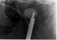
Ms E had a preoperative KUB x-ray, which identified two calculi--one measuring 11 mm and the other measuring 6 mm. Ms E was anesthetized, positioned, prepped, and draped in the usual manner. The urologist inserted a guidewire into her left ureter and used a balloon dilator to allow insertion of a rigid ureteroscope.
The urologist believed the calculi were too large to remove with stone basket forceps. He used a disposable, bare laser fiber, called a fibertome. He set the laser at 0.5 joules, 5 Hz, 2.5 watts. A total of .91 kilojoules of energy was used. The procedure took approximately 45 minutes; actual laser time was 20 minutes (intermittent). Both calculi were broken into fragments, which Ms E passed in her urine during the next several days. The urologist removed her ureteral stent in his office 48 hours after the procedure. There were no complications. On her three-month postoperative visit, Ms E remained healthy and free of cystine calculi.
CONCLUSION
The holmium:YAG laser has many applications. The value of this laser in urologic procedures to remove cystine calculi is dependent on the urologists' and perioperative staff members' training. An awareness of the holmium:YAG laser's potential for causing ureteral injuries and taking the necessary steps to avoid them is essential. Not all physicians use the holmium:YAG laser because of the potential risk for ureteral injuries; however, for patients who suffer with cystine calculi, holmium:YAG ureterolithotripsy offers an alternative to open urologic procedures. Laser treatments are cost-effective, less invasive, and highly successful.
Denise H. Adams, RN, BSN, CNOR, is a laser program specialist, Lutheran Medical Center, Wheat Ridge, Colo.
Brett B. Abernathy, MD, is a urologist in private practice, Wheat Ridge, Colo.
NOTES
(1.) D Smith, E A Tanagho, J W McAninch, Smith's General Urology, 13th ed (Norwalk, Conn: Appleton & Lange, 1992) 282.
(2.) Ibid, 283.
(3.) P C Walsh et al, eds, Campbell's Urology, sixth ed (Philadelphia: W B Saunders, 1992) 2117.
(4.) Smith, Tanagho, McAninch, Smith's General Urology, 13th ed, 283.
(5.) Ibid.
(6.) J Y Gillenwater et al, Adult and Pediatric Urology, second ed (St Louis: Mosby-Year Book, 1991) 1981.
(7.) A Smith, B S Stien, R C Benson Lasers in Urologic Surgery, third ed (St Louis: Mosby-Year Book, 1994) 190.
(8.) Ibid, 192.
(9.) Ibid.
(10.) S Baba, H Asanuma, H Tazaki, "Pulsed dye laser lithotripsy for ureteral stone fragmentation," Keio Journal of Medicine 42 (December 1993) 209-211.
(11.) Association of Operating Room Nurses, Inc, "Recommended practices for laser safety in the practice setting," in AORN Standards and Recommended Practices (Denver: Association of Operating Room Nurses, Inc, 1996) 211-215.
New Model Aids Understanding of Digestion
A July 1996 news release from Rush-PresbyterianSt Luke's Medical Center, Chicago, announces that a live rat model developed by Rush neonatologists is helping scientists who study malfunctions of the small intestine understand exactly how humans absorb what they eat. The model mirrors human absorption of nutrients; therefore, the live rat model is physiologically more accurate than previous research methods that used tissue slices and intestines exposed to surgical conditions. The model, the release states, is revolutionizing research into diseases that affect digestion, such as the intestinal problems suffered by many patients with cystic fibrosis. The model also can help scientists understand the intestine's interaction with the liver, which helps digest and store nutrients.
New Model Aids Understanding of Digestion (news release, Chicago: Rush-Presbyterian-St Luke's Medical Center, July 1996) 2.
COPYRIGHT 1996 Association of Operating Room Nurses, Inc.
COPYRIGHT 2004 Gale Group


 Home
Home A
A Gastroesophageal reflux...