Giuseppe Oreto, M.D.,(*1) Antonino Donato, M.D.;(*2) David Blumsohn, M.B., F.C.C.P.;([unkeyable]) Keith Segal, M.B.;([unkeyable]) Neil Maresky, M.B.;([unkeyable]) Leo Schamroth, M.D.([unkeyable])
We present a case of atrioventricular (AV) junctional parasystole manifesting with ventricular fusion beats due to the presence of an accessory AV conduction pathway. Ventricular fusion beats are usually impossible in AV junctional parasystole. In the reported case the ventricular fusion occurs because the ectopic AV junctional impulse is conducted through the His bundle, whereas the sinus impulse is conducted to the ventricles through the Kent bundle.
An AV junctional parasystole is the expression of a protected, and thus independent, pacemaker that is situated within the AV junction. The arrhythmia is usually diagnosed in the presence of: (a) premature ventricular complexes of AV junctional origin, which manifest with variable coupling intervals; (b) mathematically related interectopic intervals; and (c) absence of ventricular fusion beats.[1] This report deals with a case of AV junctional parasystole where ventricular fusion beats do occur. This is due to the concomitant presence of an accessory AV conduction pathway.
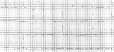
The ECG (Fig 1, selected strips of standard leads 1, 2, and 3) was recorded from a 40-year-old man affected by mild hypertension. The following is evident: (1) The basic rhythm is sinus bradycardia, at a rate ranging from 42 to 54. (2) The P-R interval of conducted sinus beats is 0.11 s. A marked initial slurring of the R wave, a delta wave, is also evident in lead 2. The QRS duration of sinus beats is 0.11 s. (3) There are several QRS complexes of ectopic origin. They differ in configuration from the basic sinus complexes. The ectopic beats reflect the following features that are absent in the sinus beats: (a) small q waves in leads 2 and 3; (b) small s waves in lead 1. The frontal plane QRS axis of ectopic complexes is +60 [degrees]. This is very different from the QRS axis of conducted sinus complexes, which is about 0 [degree]. Furthermore, no delta wave is evident in the ectopic complexes, the duration of which is 0.09 s. (4) The ectopic beats are at times dissociated from the near-synchronous sinus P waves (for example, the second QRS complex in the top strip), whereas in other instances a retrograde P' wave follows the premature beats, being partially hidden in the QRS complex (for example, the penultimate beat in the second strip). (5) Several fusion beats, labeled F, are also evident in lead 3. They reflect an intermediate configuration between the "pure" ectopic complexes and the sinus complexes. (6) The coupling intervals of the ectopic beats are variable, and range from 1.03 to 1.42 s. The interectopic intervals are mathematically related: the short intervals range from 2.40 to 2.45 s, while the long intervals are multiples of 2.37 to 2.50 s.
DISCUSSION
A form of AV junctional parasystole, associated with the Wolff-Parkinson-White (WPW) syndrome, is evident from this tracing. The AV junctional origin of the ectopic rhythm results from the narrow configuration and normal duration of the ectopic complexes. It is evident that the impulse delivered from the AV junctional focus has no access to the Kent bundle, being conducted to the ventricles by the normal AV pathway only. This accounts for the difference between the sinus and the ectopic QRS complexes: the sinus beats resulting, in fact, from a fusion between the impulse simultaneously conducted by the Kent bundle and the normal AV pathway.
The parasystolic mechanism is evident, since the coupling intervals of ectopic beats are variable, and the interectopic intervals are mathematically related. The rather uncommon finding in this tracing is the presence of ventricular fusion beats, which result from simultaneous ventricular activation by both the sinus and the parasystolic impulse. Ventricular fusion beats are impossible in uncomplicated AV junctional parasystole, since the conduction to the ventricles of both sinus and parasystolic impulses depends on a single pathway.[1] Therefore, whenever this pathway (the AV node and the His bundle) is entered and depolarized by one (sinus or ectopic) impulse, a near-synchronous impulse delivered by the other pacemaker cannot be conducted. Furthermore, if the two impulses would succeed in entering the AV node simultaneously, they would necessarily merge within the conduction pathway, so that the final result would be a normal QRS complex, and never a fusion beat.
It is worth noting, however, that atrial fusion beats are still possible in AV junctional parasystole. In the present case, ventricular fusion beats occur whenever a sinus P wave immediately precedes a parastystolic complex. The sinus impulse, however, is conducted to the ventricles by the Kent bundle only (Fig 2).
(*) Istituto di Clinica Medica e Terapia Medica, Cattedra di Malattie Cardiovascolari, Universita di Messina, Messina, Italy.
([unkeyable]) Baragwanath Hospital and University of the Witwatersrand. Johannesburg, South Africa.
REFERENCE
1 Schamroth L. The disorders of cardiac rhythm. 2nd ed. Oxford: Blackwell Scientific Publications, 1980
COPYRIGHT 1989 American College of Chest Physicians
COPYRIGHT 2004 Gale Group


