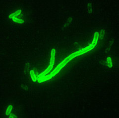
Real-time fluorescence polymerase chain reaction is a microbial identification method that can provide rapid and accurate results using a field-deployable thermocycler, the RAPID ("ruggedized" advanced pathogen identification device). A Yersinia pestis-specific TaqMan assay required approximately 75 minutes and achieved a sensitivity of 100 fg of Y. pestis genomic DNA (20 genome equivalents). Specificity testing against a genomic DNA cross-reaction panel comprised of 22 bacterial species encountered in the respiratory tract resulted in no false positives. No cross-reaction occurred with human genomic DNA.
Introduction
Quick and accurate identification assays are crucial in response to biological agents in warfare and bioterrorist ats. Biological agents that are currently of the greatest threat are Bacillus anthracis, variola major (smallpox virus), and Yersinia pestis.1 The plague bacillus, Y. pestis, has an ominous history. During three major pandemics over the past 1,500 years, it is estimated to have caused 100 to 200 million deaths with mortality rates as high as 50% to 60% during several epidemics.2 During a recent plague outbreak in Surat, India, dozens of deaths from the bacillus were confirmed by serology; however, the loss of life and suffering attributed to medical facilities overwhelmed by a panic-stricken population will never be known.3 The economic loss was in the billions. The threat as a biological weapon is significant; it is estimated that more than 50% of infected individuals will die without early antibiotic therapy, and the potential societal impact is staggering.4-6 Quick and accurate identification assays ensure implementation of antibiotic therapy and countermeasure responses in a time-critical manner as well as facilitate the dissemination of public awareness information to assuage alarm.
Molecular assays that can be implemented in the field are inherently valuable because identification can be achieved with enhanced sensitivity and specificity and identification is not diminished with nonviable organisms.7 A collaborating laboratory provided primer and fluorogenic TaqMan hybridization probe sequences designed to establish a high throughput polymerase chain reaction (PCR) assay for the identification of Y. pestis. Although the high throughput assay system achieved excellent sensitivity and specificity, it does not allow for rapid identification in a field environment. Our laboratory has adapted the high throughput TaqMan assay to a field-deployable assay platform, the RAPID ("ruggedized" advanced pathogen identification device).8-11 The TaqMan probe is a dual fluorophore-labeled, oligonucleotide specific to the PCR product, amplified Y. pestis DNA, generated by primer extension of the organism's DNA template.12 Increasing fluorescence represents the quantity of DNA target sequence amplified over the duration of the PCR. By implementing a standard curve, linear regression analysis quantifies the number of DNA target sequences in an unknown sample.
The time required for a conventional laboratory analysis is unacceptable in situations in which a biological weapon has been used against fielded troops or in any bioterrorism situation. The need for accurate in situ identification systems for biowarfare agents is paramount.13 This article describes a highly sensitive and specific, real-time, fluorogenic PCR assay for the detection of Y. pestis using a field-deployable thermocycler.
Materials and Methods
Assay sensitivity was determined with Y. pestis genomic DNA obtained from a collaborating laboratory. Identification of the strain is undisclosed. Specificity testing was conducted with human genomic DNA (Roche Molecular Biochemicals, Mannheim, Germany) and genomic DNA was isolated from bacterial cultures prepared from laboratory stocks (Clinical Microbiology Laboratory, Air Force Institute for Environment, Safety, and Occupational Health Risk Analysis, Epidemiology Surveillance Division, Brooks Air Force Base, Texas). Genomic DNA was isolated from overnight cultures using the MagNA Pure LC System and MagNA Pure LC Total Nucleic Acid Isolation kit (Roche Diagnostics, Mannheim, Germany).14,15
Bacterial organisms commonly cultured from the respiratory tract were selected and recovered from frozen cultures, restreaked to single colonies on agar plates, cultured under conditions appropriate for each species, and identification confirmed with conventional biochemical analyses and the Vitek Automicrobic system (Biomerieux-Vitek, Hazelwood, Missouri). A single colony of each organism was transferred with a sterile loop to liquid culture media, was incubated for 18 to 48 hours, and 200 [mu]L of each liquid culture was pipetted into the MagNA Pure LC sample cartridge. All postloading processing was completed in a closed system by automated robotics with preformatted reagents and a nucleic acid isolation matrix. Cell lysis and nucleic acid stabilization was completed with buffer containing guanidinium thiocyanate and proteinase K. Nucleic acid bound to the surface of magnetic glass particles was isolated from other cellular components by washing and eluting with a low-salt buffer.
Preliminary assay optimization was performed on a LightCycler (Roche Molecular Biochemicals) and transferred to the RAPID (Idaho Technology, Salt Lake City, Utah). Primer and TaqMan probe target sequences were provided by a collaborating laboratory. Identification of primer and probe sequences and target locus is undisclosed. All reagents were purchased commercially (QuantiTect Probe PCR kit, Qiagen, Valencia, California) containing 2x concentrations of HotStarTaq DNA polymerase, QuantiTech probe PCR buffer, dNTP mix including dUTP, and 8 mM MgCl^sub 2^. Forward and reverse primer final concentrations were optimized at 0.50 [mu]M each and the TaqMan probe concentration at 0.03 [mu]M. Assays were carried out in sealed LightCycler capillary tubes with 20 [mu]L of final volume of reaction mixture comprised of the above master mix, primers, probe, and DNA template. Assay conditions were optimized to a single initial denaturation cycle of 15 minutes at 94[degrees]C followed by 45 cycles of denaturation at 94[degrees]C for 0 seconds and extension at 60[degrees]C for 60 seconds with the single fluorescent data acquisition step after each cycle. The concentration of Y. pestis control DNA was determined spectrophotometrically, and a logarithmic dilution series was constructed ranging from 10.0 ng to 1.0 fg or 2.1 e6 to 2.1 e1 genome equivalents, respectively, per 20 [mu]L of reaction volume. No-template controls were prepared by adding a 2-[mu]L volume of PCR grade water in 18 [mu]L of master mix. The concentration of each cross-reaction microbial organisms was standardized at 1.0 ng of DNA per 20 [mu]L of reaction volume.
Results
Sensitivity of the PCR In Vitro Assay
The sensitivity of the PCR assay was optimized to a minimum limit of detection of 100 fg of purified Y. pestis genomic DNA (20 genome equivalents). Real-time PCR assay sensitivity optimization resulted in a linear regression curve across a dilution series of 10 ng to 100.0 fg of Y. pestis genomic DNA (2.1 e6 to 2.1 e1 genome equivalents) with an error of
Specificity of PCR In Vitro Assay
Specificity of the assay when tested with 1.0 ng of 22 cross-reactivity panel bacterial organisms and 14.0 ng (4.4 e3 genomic equivalents) of human DNA displayed no detectable fluorescence above background (Table I). Spiked cross-reaction panel data verified the absence of PCR inhibition (Table I). When spiked with 2. 1e5 genomic equivalents (1.0 ng) of Y. pestis DNA, the average threshold of fluorescence detection or critical threshold (C1) was ~17 (N = 23; mean, 16.99; range, 15.73-21.67). Postoptimization testing under identical conditions with 2.1e5 genomic equivalents (1.0 ng) of Y. pestis DNA had an average C^sub t^ of ~20 (JV = 10; mean, 19.98; range, 18.10-21.69).
During cross-reaction testing initial fluorescence data reported two false positives, Pseudomonas aeruginosa (C^sub t^ = 41.64; calculated DNA quantity = 5.6 e to 6 ng) and Staphylococcus aureus (C^sub t^ = 40.86; calculated DNA quantity = 8.3 e to 6 ng). However, when the standard curve was fit to
Discussion
This study shows that real-time fluorescence PCR using field-deployable instrumentation and preformatted reagents provides a sensitive and specific assay for identification of Y. pestis DNA. Testing demonstrated a range of detection from 10.0 ng to 100.0 fg of Y. pestis genomic DNA or 2.1 e6 to 2.1 e1 genome equivalents, respectively, and a specificity of 100% (24/24) across a genomic DNA panel of Y. pestis, human, and purified DNA from 22 bacterial organisms encountered in the respiratory tract. There was no indication of inhibition of the PCR in any of the cross-reaction panels or human genomic DNA samples. Spiked cross-reaction panels and human genomic DNA samples reported fluorescence nominally when exposed to the PCR. The inherent value of the assay is in its adaptability to a field-deployable assay platform and potential for direct detection from blood and oropharyngeal specimens in the field. The RAPID has a 32-sample capacity and achieves high-speed thermal cycling through fan-driven air rather than heat block conduction. In this study, assay results were obtained in approximately 75 minutes, were highly reproducible, and the risk of sample cross-contamination was greatly reduced due to the closed capillary component of the system. With the exception of reagent preparation, the assay itself is completely automated.
However, microbiological pathogen detection technology, including field-deployable platforms, has outpaced the development of field-deployable nucleic acid extraction and stabilization technologies. Isolation of a DNA template of sufficient purity for the PCR with traditional laboratory technology typically requires 1 hour or more of labor-intensive activities requiring unwieldy equipment and multiple reagents.16 Nucleic acid purification reagents include an assortment of hazardous materials such as TRIzol, chloroform, isopropanol, and ethanol that complicate logistics exponentially. The development of simple, robust, and rapid technologies to efficiently isolate and stabilize nucleic acid in the field and that are easily transported is currently a limiting constraint in the realization of an omnipotent field assay platform. In our laboratory, we have adapted a solid-phase DNA purification system that has proven a simple and efficient method for rapid DNA purification and confirmation testing from Streptococcus pneumoniae isolates.17 We have recently implemented the solid-phase DNA purification method to identify S. pneumoniae in pleura fluid and tissue from patient samples (M.D. Poulter, J.D. Cherry, J.G. Deville, et al., manuscript in preparation). We have also successfully implemented the method to isolate and identify Salmonella DNA from food samples18 and stool (M.D. Poulter, J.D. Cherry, J.G. Deville, et al., manuscript in preparation) as well as Bacillus anthracis from body fluids of cattle.19 We successfully field tested the method with human-deposited microorganisms in Arctic soil samples, NASA Haughton-Mars Project, Devon Island, Canada, 2001 field season (R.M. Roudabush, L.T. Daum, K.L. Lohman, and J.C. McAvin, unpublished data). We plan to test this method for the isolation of Y. pestis DNA from soil as well as blood and oropharyngeal samples. The next level of nucleic acid purification and stabilization technology will be more efficiently advanced and protocols standardized through cooperative military, academic, and corporate partnership.
Real-time identification of biological agents used in warfare and bioterrorism will allow military commanders and homeland defense agencies to obtain vital information on conditions in the quickest possible time and direct the implementation of appropriate force protection countermeasures and medical responses. From continued development of field assay systems for detection of biological agents of immediate threat will evolve research and development paradigms for real-time multiplex PCR assays for simultaneous detection of multiple biowarfare agents20,21 and DNA microarray technologies22 that will meet the challenge of forthcoming genetically engineered, supervirulent bacteria and viruses.
Acknowledgments
We thank Kathy Beninga and Glen Jackson for their technical assistance and Rebecca Medina for help in preparing this manuscript. Thank you to Dr. David Hale (Department of Biology, U.S. Air Force Academy) for comments and support.
References
1. Polgreen PM, Helms C: Vaccines, biological warfare, and bioterrorism. Prim Care 2001; 28: 807-21.
2. Perry RD, Fetherston JD: Yersinia pestis: etiologic agent of plague. Clin Microbiol Rev 1997; 10: 35-66.
3. Putzker M, Sauer H, Sobe D: Plague and other human infections cause by Yersinia species. Clin Lab 2001; 47: 453-66.
4. Broussard LA: Biological agents: weapons of warfare and bioterrorism. Mol Diagn 2001; 6: 323-33.
5. Inglesby TV, Dennis DT, Henderson DA, Barlett JG, Ascher MS, Eitzen E, et al: Plague as a biological weapon: medical and public health management: working group on civilian biodefense. JAMA 2000; 283: 2281-90.
6. Olive MD, Bean P: Principles of applications of methods tor DNA-based typing of microbial organisms. J Clin Microbiol 1999; 37: 1661-9.
7. Niemeyer DM: Polymerase chain reaction: a link to (he future. Milit Med 1998; 163: 226-8.
8. Niemeyer DM, Jaffe RI, Wiggins LB: Feasibility determination for use of polymerasc chain reaction in the U.S. Air Force air-transportable hospital field environment: lessons learned. Milit Med 2000; 165: 816-20.
9. Wittwer CT, Ririe KM, Andre RV, David DA, Gundry RA, Balis UJ: The lightcycler: a microvolume multisample fluorimeter with rapid temperature control. BioTechniques 1997; 22: 176-81.
10. Wilson JR: Chem-detection. Milit Med Technol 2001; 5: 20-2.
11. Wittwer CT, Herrmann MG, Moss AA, Rasmussen RP: Continuous fluorescence monitoring of rapid cycle DNA amplification. BioTechniques 1997; 22: 130-8.
12. Enserink M: Anthrax: biodefense hampered by inadequate tests. Science 2001; 294: 1266-7.
13. Kessler HH, Muhlbauer G, Stelzl E, Daghofer E, Santner BI, Marth E: Fully automated nucleic acid extraction: MagNA Pure LC. Clin Chem 2001; 47: 1124-6.
14. Costa C, Costa JM, Desterke C, Botterel F, Cordonnier C, Bretagne S: Real-time PCR coupled with automated DNA extraction and detection of galactomannan antigen in serum by enzyme-linked immunosorbent assay for diagnosis of invasive aspergillosis. J Clin Microbiol 2002; 40: 2224-7.
15. Higuchi R: Simple and rapid preparation of samples for PCR. PCR Technology: Principles and Applications for DNA Amplification, pp 31-8. New York, Stockton Press, 1989.
16. McAvin JC, Reilly PA, Roudabush RM, Barnes WJ, Salmen A, Jackson GW, et al: Sensitive and specific method for rapid identification of Streptococcus pneumoniae using real-time fluorescence PCR. J Clin Microbiol 2001; 39: 3446-51.
17. Daum LT, Barnes WJ, McAvin JC, Neidert MS, Cooper LA, Huff WB, et al: Real-time PCR dection of salmonella in suspected foods from a gastroenteritis outbreak in Kerr County, Texas. J Clin Microbiol 2002; 40: 1-3.
18. Cooper LA, Atchley DH, Hadfield TL, Kelly JM, Ashibrd D, Neidert MS, et al: Detection of Bacillus anthracis in veterinary clinical samples using field deployable real-time iluorogenic PCR. Presented at the International Conference on Emerging Infectious Diseases, ICEID, 2002.
19. McDonald R, Cao T, Borschel R: Multiplexing for the detection of multiple biowarfare agents shows promise in the field. Milit Med 2001; 166: 237-9.
20. Wittwer CT, Herrmann MG, Gundry CN, Elenitoba-Johnson KS: Real-time multiplex PCR assays. Methods 2001; 25: 430-42.
21. Brown PO, Botstein D: Exploring the new world of the genome with DNA microarrays. Nat Genet 1999; 21(Suppl 1): 33-7.
22. Ye RW, Wang T, Bedzyk L, Croker KM: Applications of DNA microarrays in microbial systems. J Microbiol Methods 2001; 47: 257-72.
Guarantor: James C. McAvin, MS
Contributors: James C. McAvin, MS*[dagger]; Mariana A. McConathy, MS*; Andrew J. Rohrer, BS[double dagger]; Lt Col William B. Huff, BSC USAF*; Maj William J. Barnes, BSC USAF*; Kenton L. Lohman, PhD*
* Epidemlological Surveillance Division, Molecular Epidemiology Branch, Air Force Institute for Environment, Safety, and Occupational Health Risk Analysis, Brooks Air Force Base, San Antonio, TX 78235-5237.
[dagger] Molecular Epidemiology, AFIERA, 2730 Louis Bauer Drive, Building 930, Brooks Air Force Base, San Antonio, TX 78235-5237.
[double dagger] United States Air Force Academy, Colorado Springs, Colorado.
This manuscript was received for review in October 2002 and accepted for publication in December 2002.
Copyright Association of Military Surgeons of the United States Oct 2003
Provided by ProQuest Information and Learning Company. All rights Reserved


