Abstract
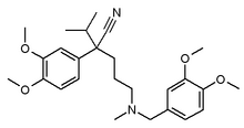
Calcium-channel blockers (CCB), including verapamil, nifedipine, and diltiazem, are one of the most widely prescribed class of drugs in the treatment of cardiovascular disease. In the last several years, CCBs have been linked with a distinct cutaneous subset in the lupus spectrum, subacute cutaneous lupus erythematosus (SCLE), and we describe a case induced by verapamil.
**********
Case Report
A 56-year-old African-American female presented with a 1-month history of a progressively worsening pruritic eruption that began on the lower extremities and spread to her arms, abdomen, and back. There were no clear exacerbating factors and she received no prior treatment.
The patient's past medical history is significant for hepatic cirrhosis secondary to alcohol abuse, esophageal varices, hypertension, and surgical lysis of peritoneal adhesions. She had no personal or family history of skin disease or photosensitivity. Her only medications were verapamil X 6 years and aspirin. She had no known drug allergies.
Physical examination revealed hyperpigmented, hyperkeratotic papules and plaques with active erythematous borders over the lower extremities (Figure 1), abdomen, back, and arms. Examination of scalp, face, ears, neck, arms, palms, and soles were unremarkable. Oral mucous membranes revealed hyperpigmentation, but no inflammatory lesions or lacy white pattern of buccal mucosa. Her sclerae were icteric.
Histology sections of lesional skin from the lower leg and abdomen showed marked hyperkeratosis and prominent follicular plugging. Necrotic keratinocytes, vacuolization of the basal layer, and focally thickened basement membrane were seen within an atrophic epidermis (Figure 2). There was a perivascular and perifollicular infiltrate of lymphocytes and histiocytes in the dermis. Mucin deposition was focally noted. Direct immunofluorescence examination demonstrated weak, granular, and linear deposition of IgG at the dermal epidermal junction. Examination for IgM, IgA, and C3 deposition was negative. Potassium hydroxide preparation was negative for fungal hyphae.
Laboratory studies demonstrated a positive antinuclear antibody (ANA) titer of 1:160 in a nucleolar pattern, and positive anti-Ro/anti-La antibodies. Antibodies to dsDNA, Jo-1, U1-RNP, scl-70, and Smith were not detected.
The patient's findings were felt to be consistent with subacute cutaneous lupus erythematosus (SCLE). The verapamil was discontinued, and the patient was treated topically with fluocinonide 0.05% ointment. On the 2-week follow-up visit, the lower extremity and abdominal lesions were less erythematous and scaly, but hyperpigmentation, without scarring occurred. The patient had developed new 4 to 5 mm violaceous lichenoid lesions of the upper extremities and a new plaque on the abdomen. Two months after initial examination she had only post-inflammatory hyperpigmentation.
[FIGURE 1 OMITTED]
Discussion
In 1997, Crowson and Magro (1) reported a series of 9 patients who experienced an adverse cutaneous reaction to CCBs, which suggested a clinical diagnosis of SCLE. Clinically, these patients exhibited annular or papulosquamous eruptions without evidence of arthritis, central nervous system, or renal involvement. Most cases had a history of photosensitivity and the rash is photo-distributed. Serologic studies showed all but 2 of the patients were ANA positive and all but one expressed anti-Ro and/or anti-La antibodies. In all cases the histopathology and immunofluorescence (IF) findings were consistent with SCLE. Of the 9 patients, 4 had been taking diltiazem, 4 were on verapamil, and 1 was on nifedipine. None had a prior history of a similar eruption or of positive serology for lupus erythematosus. The medications were discontinued with complete resolution of skin eruptions within 2 to 12 weeks of drug cessation. An additional case of nifedipine induced SCLE was reported recently. (2) Srivastava et al (3) reviewed retrospectively 120 patients with positive anti-Ro/SSA antibodies and found 70 had histology confirmed cutaneous lupus erythematosus. Fifteen had newly started drugs associated with SCLE within 6 months, including 3 patients on CCB. A new calcium channel blocker, nitrendipine has also been reported to cause SCLE. (4)
We believe that our case adds further support to a growing body of evidence that calcium channel blockers can be directly linked to the induction of SCLE. Other drugs have been shown to induce SCLE including terbinafine, (5,6) hydrochlorthiazide, (7) angiotensin-converting enzyme inhibitors, (3) statins, (3) interferons, and griseofulvin. (8)
In conclusion, though cutaneous adverse reactions to calcium channel blockers are uncommon, (9) clinicians should nonetheless be aware of its association with SCLE. Drug-induced SCLE is reversible, but is often unrecognized. (10) Treatment includes the use of topical steroids and cessation of the causative agent. Patients can experience the eruption for up to 3 months after cessation of the offending drug.
[FIGURE 2 OMITTED]
References
1. Crowson AN, Magro CM. Subacute cutaneous lupus erythematosus arising in the setting of calcium channel blocker therapy. Hum Pathol. 1997;28:67-73.
2. Gubinelli E, Cocuroccia B, Girolomoni G. Subacute cutaneous lupus erythematosus induced by nifedipine. J Cutan Med Surg. 2003;7:243-246.
3. Srivastava M, et al. Drug-induced Ro/SSA-positive cutaneous lupus erythematosus. Arch Dermatol. 2003;139:45-49.
4. Marazo AV, et al. Nitrendipine-induced subacute cutaneous lupus erythematosus. Euro J Dermatol. 2003;13:213-6.
5. Bonsmann G, et al. Terbinafine-induced subacute cutaneous lupus erythematosus. J Am Acad Dermatol. 2001;44:925-931.
6. Callen JP, Hughes AP, Culp-Shorten C. Subacute cutaneous lupus erythematosus induced or exacerbated by terbinafine: a report of 5 cases. Arch Dermatol. 2001;137:1196-8.
7. Parodi A, Romagnoli M, Rebora A. Subacute cutaneous lupus erythematosus-like eruption caused by hydrochlorothiazide. Photodermatology. 1989;6:100-102.
8. Miyagawa S, et al. Subacute cutaneous lupus erythematosus lesions precipitated by griseofulvin. J Am Acad Dermatol. 1989;21:343-346.
9. Stern R, Khalsa JH. Cutaneous adverse reactions associated with calcium channel blockers. Arch Intern Med. 1989;149:829-832.
10. Callen JP. Drug-induced cutaneous lupus erythematosus, a distinct syndrome that is frequently unrecognized. J Amer Acad Dermatol 2001;45:315-6.
Boaz Kurtis MD, (a) Matthew J. Larson MD, (b) Mai P. Hoang MD, (c) Jack B. Cohen DO (c)
a. Department of Medicine, St. Luke's-Roosevelt Hospital Center, New York, NY
b. Columbia Park Medical Group, Brooklyn Park, MN
c. University of Texas Southwestern, Dallas, TX
Address for Correspondence
Jack B. Cohen DO
Department of Dermatology
University of Texas Southwestern
5323 Harry Hines Blvd.
Dallas, TX 75390-9190
e-mail: jack.cohen@utsouthwestern.edu
Phone: 214-648-5770
COPYRIGHT 2005 Journal of Drugs in Dermatology, Inc.
COPYRIGHT 2005 Gale Group


