Mitral valve endocarditis typically results in mitral regurgitation. However, endocarditis leading to functional mitral stenosis is uncommon and, when present, fungal organisms are typically implicated. Thus, obstructive-type bacterial endocarditis due to large vegetations blocking the mitral valve orifice is a rare occurrence, with approximately 20 reported cases in the literature. We report on two patients with bacterial endocarditis and severe functional mitral stenosis requiring emergent surgery. Additionally, this is the first report of vancomycin-resistant enterococcus causing endocarditis and functional mitral stenosis. The discussion emphasizes the hemodynamic instability of these patients and need for early surgical intervention.
Key words: bacterial endocarditis; echocardiography; enterococcal infection; mitral valve stenosis; vegetations
Abbreviations: MRSA = methicillin-resistant Staphylococcus aureus; TEE = transesophageal echocardiography; VRE = vancomycin-resistant enterococcus
**********
Valvular dysfunction in infective endocarditis implies the development of a new regurgitant lesion. The occurrence of an obstructive or "functionally" stenotic valvular condition in the setting of endocarditis is rare and often goes clinically unsuspected until late in the course of the disease. (1-8) Moreover, suspicion of this condition usually surfaces only in cases of fungal endocarditis. (9,10) Importantly, when present, the management of such a scenario is medically challenging and uniformly warrants emergent surgery. We describe two cases of bacterial endocarditis presenting with functional mitral stenosis due to obstructive vegetations at the mitral valve orifice. Novel to our report are the microorganisms involved in this "obstructive-type" of endocarditis, namely methicillin-resistant Staphylococcus aureus (MRSA) and vancomycin-resistant enterococcus (VRE). A discussion on the management of these patients and a brief review of the literature is provided.
CASE REPORTS
Case 1
A 54-year-old African-American woman presented with fevers, respiratory distress, and mental status changes. Her medical history was significant for end-stage renal disease with secondary hyperparathyroidism, long-standing hypertension, and IV drug abuse. The week prior to hospital admission, she missed several sessions of hemodialysis, becoming progressively lethargic, and was brought by her family to the emergency department. On presentation, her temperature was 39.3[degrees]C, BP was 97/65 mm Hg, and heart rate was 120 beats/min. Physical examination was remarkable for jugular venous distension, an apical holosystolic murmur that radiated to the axilla, a short apical diastolic murmur, and bilateral pulmonary crackles up to the mid-lung fields.
She underwent urgent hemodialysis, after which blood culture specimens were collected and empiric IV antibiotics initiated. A transthoracic echocardiogram was obtained that revealed severe left ventricular hypertrophy with hyperdynamic systolic function and cavity obliteration, severe right ventricular dysfunction with an estimated systolic pressure of 80 mm Hg, and mitral annular calcification with severe mitral regurgitation. Additionally, an ill-defined large echogenic mass was noted in the left atrium. By conventional Doppler velocity measurements across the mitral inflow, a significant diastolic transmitral gradient was detected, suggestive of severe mitral stenosis. Subsequently two sets of blood cultures grew MRSA. At this point, the diagnosis of infective endocarditis was established, and transfer to our facility for further management was arranged.
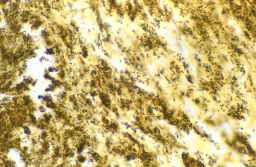
Shortly after her arrival, she was intubated and a pulmonary artery catheter was placed to assist in her management. Initial pulmonary artery pressures were 96/55 mm Hg, with similar concomitant systemic pressures and a low cardiac index despite optimal medical therapy. Given her tenuous hemodynamic status, transesophageal echocardiography (TEE) was performed to better define any potential endocarditis-related mechanical complications. TEE confirmed the presence of severe mitral regurgitation and severe functional mitral stenosis secondary to a large, mobile vegetation attached to the mitral annulus (Fig 1). Based on the findings and ongoing hemodynamic embarrassment, the decision was made to proceed with urgent surgery. Intraoperatively, surgical findings demonstrated a large vegetation (Fig 2) arising from the region of mitral annular calcification and a large subannular mitral abscess resulting in atrioventricular separation. She underwent debridement of the posterior mitral leaflet and the atrioventricular groove, autologous pericardial patch, repair of the separation between the left atrium and the left ventricle, and mitral valve replacement. Postoperatively, her hospital course was uneventful, with a return of pulmonary pressures returned to near-normal values. Subsequent blood culture findings remained negative. She was discharged home receiving long-term IV antibiotic therapy and has continued to do well.
[FIGURES 1-2 OMITTED]
Case 2
A 51-year-old white man presented with a 1-month history of low-grade fevers, fatigue, and nonproductive cough, with several days of increasing dyspnea and orthopnea. His medical history was significant for longstanding hypertension and peripheral vascular disease. His surgical history included mitral valve repair for myxomatous disease, complicated years later by Streptococcus viridans endocarditis, requiring a repeat operation the year prior to the current hospital admission (mitral valve replacement with a Carpentier-Edwards bioprosthesis). A left-sided empyema and VRE septic thrombophlebitis complicated his postoperative course.
On the current presentation, his physical examination revealed a chronically ill-appearing man in respiratory distress with a BP of 94/63 mm Hg and oxygen saturation of 91% on room air. Cardiac examination disclosed an elevated jugular venous pressure, prominent right ventricular heave, and wide splitting of the second heart sound with a prominent pulmonic component without any audible murmur. Pulmonary examination revealed bilateral diffuse crackles and decreased breath sounds at the left lung base. ECG showed sinus tachycardia, first-degree atrioventricular block, and nonspecific ST-T changes. The patient was started on empiric antibiotics after appropriate blood culture samples were collected, and transthoracic echocardiography was performed that showed normal left ventricular size and function, and a severely dilated and dysfunctional right ventricle, a finding not present on a prior study. Right ventricular systolic pressure was estimated at 76 mm Hg. In addition, there was moderate aortic insufficiency and a large mobile echodensity attached to the prosthetic leaflets partially occluding the mitral orifice. Follow-up TEE confirmed the extensive prosthetic echodensity, protruding into the left atrium, consistent with a large vegetation attached to the mitral prosthesis. Transmitral diastolic gradients by conventional Doppler velocity measurements were consistent with severe mitral stenosis (mean gradient 18 mm Hg). The prosthesis itself was well seated with only trivial prosthetic regurgitation. Blood culture findings, at this point, were positive for VRE in both sets.
Given the inability to medically manage the patient's hemodynamics, the decision was made to undergo surgery. Intraoperatively, he was found to have vegetations on the aortic valve, a small periaortic abscess, and a massive vegetation on the bioprosthetic mitral valve. Mitral and aortic valves were replaced, each with a Carpentier-Edwards bioprosthesis, and the fibrous trigone of the heart was repaired. Microbiologic examination of the explanted prosthetic mitral valve and native aortic valve confirmed the presence of VRE. Postoperatively, the patient's hemodynamics remained stable throughout his hospital stay, although right ventricular function remained severely depressed despite normalization of pulmonary pressures. Subsequent blood cultures revealed no microorganism growth, and the patient was discharged home receiving IV antibiotic therapy. The patient returned for his 1-year follow-up in overall good condition.
DISCUSSION
Mitral valve obstruction due to infective endocarditis is an uncommon occurrence. When present, suspicion of fungal endocarditis with large vegetations ("fungus balls") usually emerges, although such a scenario is commonly associated with fungal endocarditis on prosthetic heart valves. (9,10) Thus, the scenario in our cases, functional stenosis from obstructive bacterial vegetations, is quite rare. (1-8) A review of the literature from 1966 to present day using MEDLINE uncovered 20 cases of significant native mitral valve obstruction secondary to bacterial endocarditis, (6,7) with an additional 14 cases occurring in mitral prostheses. (8) Staphylococci were the most common culprit organisms. Although not uncommon with valvular prostheses, cases of fungal vegetations and obstructive native valve mitral stenosis were not found as selected reports. Our patients were infected with MRSA and VRE. To the best of our knowledge, this is the first case of bacterial endocarditis and functional mitral stenosis from obstructive vegetations involving VRE as the causative organism.
Based on prior reports, a considerable portion of cases of "obstructive-type" bacterial endocarditis have occurred in those with preexisting stenotic mitral valve disease and have eluded those with normal or purely regurgitant mitral valves, with few exceptions. (11) Various reports describe subjects with preexisting rheumatic mitral stenosis (1-4) or mitral prostheses. (8) Our first patient likely had mild underlying mitral stenosis due to significant mitral annular calcification, while the second patient had a bioprosthetic mitral valve. Comparable to the development of left atrial thrombi formation, relative mitral inflow obstruction appears to be a risk factor for the development of these large obstructive bacterial vegetations.
Consistently impressive in the management of these cases is the development of rather acute significant pulmonary hypertension and consequent right ventricular systolic failure. The inability to adequately fill the left heart coupled with a failing right heart dictate the inadequacy of medical and even mechanical (ie, intraaortic balloon counterpulsation) therapies in treating this clinical scenario. As such, patients inevitably warrant emergent surgical intervention because of significant hemodynamic instability. As supported from the available literature, overall mortality is high (6) but approaches 100% for those treated medically. (2)
In conclusion, bacterial endocarditis and functional mitral stenosis (obstructive-type endocarditis) constitutes a potentially fatal disease that requires early echocardiographic diagnosis and emergent surgical intervention. However, with modern day medical and surgical management, many patients with this condition should survive. With some exceptions, cases seem to occur in conjunction with preexisting stenotic mitral valve disease or mitral bioprosthesis. A bacterial etiology, in addition to fungal, should be included in the differential of this clinical scenario, and knowledge of the limitations of medical therapy and urgency of early surgery are critical.
REFERENCES
(1) Roberts WC, Ewy GA, Glancy DL, et al. Valvular stenosis produced by active infective endocarditis. Circulation 1967; 36:449-451
(2) Reeve R, Reeve JS, Matula G, et al. Mitral obstruction by vegetations of staphylococcal endocarditis. JAMA 1974; 228:75
(3) Matula G, Karpman LS, Frank S, et al. Mitral obstruction from staphylococcal endocarditis, corrected surgically. JAMA 1975; 233:58-59
(4) Copeland JG, Salomon NW, Stinson EB, et al. Acute mitral valvular obstruction from infective endocarditis: echocardiographic diagnosis and report of the second successfully treated case. Thorac Cardiovasc Surg 1979; 78:128-130
(5) Waller BF, McManus BM, Roberts WC. Mitral valve stenosis produced by or worsened by active bacterial endocarditis. Chest 1982; 82:498-500
(6) Ghosh PK, Miller HI, Vidne BA. Mitral obstruction in bacterial endocarditis. Br Heart J 1985; 53:341-344
(7) Prasad TR, Valiathan MS, Venkitachalam CG, et al. Unusual manifestation of valvular vegetations. Thorac Cardiovasc Surg 1988; 36:170-171
(8) Citrin BS, Mensah GA, Byrd BF. Functional mitral stenosis resulting from a large mitral valve prosthesis vegetation. South Med J 1997; 90:231-233
(9) Seelig MS, Speth CP, Kozinn PJ, et al. Patterns of Candida endocarditis following cardiac surgery: importance of early diagnosis and therapy (an analysis of 91 cases). Prog Cardiovasc Dis 1974; 17:125-160
(10) Melgar GR, Nasser RM, Gordon SM, et al. Fungal prosthetic valve endocarditis in 16 patients: an 11-year experience in a tertiary care hospital. Medicine (Baltimore) 1997; 76:94-103
(11) Enia F, Lo Mauro R, Matassa C, et al. [Acute mitral obstruction during infectious endocarditis]. G Ital Cardiol 1983; 13:347-350
* From the Department of Cardiovascular Medicine (Drs. Novaro, Jefferson, Monson, and Penn), Internal Medicine (Dr. Tiong), and Cardiothoracic Surgery (Dr. Smedira), The Cleveland Clinic Foundation, Cleveland, OH.
Manuscript received March 21, 2002; revision accepted June 21, 2002.
Correspondence to: Marc S. Penn, MD, PhD, Departments of Cardiovascular Medicine and Cell Biology, NC10, Cleveland Clinic Foundation, 9500 Euclid Ave, Cleveland, OH 44195; e-mail: pennm@ccf.org
COPYRIGHT 2002 American College of Chest Physicians
COPYRIGHT 2003 Gale Group


