Basal cell carcinoma
Basal cell carcinoma (BCC) is the most common skin cancer. It can be destructive and disfiguring. Risk is increased for individuals with a family history of the disease and a high cumulative exposure to UV light via sunlight or, in the past, carcinogenic chemicals especially arsenic. Treatment is with surgery, topical chemotherapy, x-ray, cryosurgery, photodynamic therapy, or topical immune enhancement drugs such as imiquimod. It is rarely life-threatening but if left untreated can be disfiguring, cause bleeding and produce local destruction (eg., eye, ear, nose, lip). more...
Forms
Various forms are recognised:
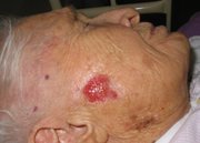
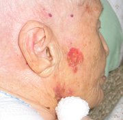
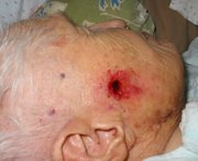
- Nodular: flesh-colored papule with telangiectasis. If it ulcerates, it becomes a "rodent ulcer" (ulcus rodens), an ulcerating nodule with (often) a pearly border.
- Cystic: rarer and hard to distinguish from the nodular form. It has a central cavity with fluid.
- Pigmented: a variant of the nodular form that may be confused with melanoma.
- Sclerosing/cicratising: a scar-like lesion.
- Superficial: a red scaling patch
About two thirds of the carcinomas occur in sun-exposed areas and onw third occur in non-sun-exposed areas, emphasizing the genetic susceptability of the basal cell cancer patients.
Diagnosis
To diagnose, a biopsy (where tissue is taken for pathological study) is done using local anesthesia. In small lesions, the tumor is generally removed in its entiriety, while larger ones are biopsied first and surgically removed later if it is confirmed that it is malignant.
Histopathology: Basal cell carcinoma is a malignant epithelial tumor arising only in skin, from the basal layer of the epidermis or of the pilosebaceous adnexa. Tumor is represented by compact areas, well delineated and invading the dermis, apparent with no connection with the epidermis. Tumor cells resemble normal basal cells (small, monomorphous) are disposed in palisade at the periphery of the tumor nests, but are spindle-shaped and irregular in the middle. Tumor clusters are separated by a reduced stroma with inflammatory infiltrate. 1
Pathophysiology
Basal cell carcinomas develop in the basal cell layer of the skin. Sunlight exposure leads to DNA crosslinking between thymidine residues. While DNA repair removes most UV-induced damage, not all crosslinks are excised. There is, therefore, cumulative DNA damage leading to mutations. Apart from the mutagenesis, sunlight depresses the local immune system, possibly decreasing immune surveillance for new tumor cells.
Prevention & Early Diagnosis
Basal cell carcinoma is the most common skin cancer. It occurs mainly in fair-skinned patients with a family history of this cancer. Sunlight is a factor in about two thirds of these cancers, but one third occur in non sun-exposed areas. Therefore, dermatologists recommend sun screens and annual skin cancer exams to prevent or provide early detection of this common tumor.
Treatment
Most basal cell carcinomas are removed surgically by dermasurgeons. A common method is "electrodessication and curettage" (ED&C). This is done by scraping the tumor out with a curette and cauterizing the base and margins. The wound is left to heal by itself (secondary intention healing). The cure rate and cosmetic result are excellent, especially in concave areas. It is also the most cost effective treatment. Surgical excision by the dermasurgeon is another option with the margins of excised tissue examined under the microscope. Certain types, like the sclerosing basal cell cancers may need a wider margin, as they develop subtle processes that project outside the visible part of the tumor.
Read more at Wikipedia.org


 Home
Home A
A Babesiosis