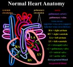
Normal heart anatomy
Find information on thousands of medical conditions and prescription drugs.

Great vessels transposition

Transposition of the great vessels (TGV) is a group of congenital heart defects (CHDs) involving an abnormal spatial arrangement of any of the primary vessels: superior and/or inferior vena cavae (SVC, IVC), pulmonary artery, pulmonary veins, and aorta. more...

Home
Diseases
A
B
C
D
E
F
G
Galactorrhea
Galactosemia
Gardner's syndrome
Gastric Dumping Syndrome
Gastroesophageal reflux
Gaucher Disease
Gaucher's disease
Gelineau disease
Genu varum
Geographic tongue
Gerstmann syndrome
Gestational trophoblastic...
Giant axonal neuropathy
Giant cell arteritis
Giardiasis
Gigantism
Gilbert's syndrome
Gilles de la Tourette's...
Gingivitis
Gitelman syndrome
Glanzmann thrombasthenia
Glioblastoma
Glioblastoma multiforme
Glioma
Glomerulonephritis
Glossodynia
Glossophobia
Glucagonoma
Glucose 6 phosphate...
Glutaryl-CoA...
Glycogen storage disease
Glycogen storage disease...
Glycogenosis type IV
Glycosuria
Goiter
Goldenhar syndrome
Goodpasture's syndrome
Graft versus host disease
Granulocytopenia
Graves' disease
Great vessels transposition
Growth hormone deficiency
Guillain-Barré syndrome
Gymnophobia
H
I
J
K
L
M
N
O
P
Q
R
S
T
U
V
W
X
Y
Z
Medicines

Description

In a normal heart, oxygen-depleted ("blue") blood is pumped from the right side of the heart, through the pulmonary artery, to the lungs where it is oxygenated. The oxygen-rich ("red") blood then returns to the left heart, via the pulmonary veins, and is pumped through the aorta to the rest of the body, including the heart muscle itself.

Transposed vessels can present a large variety of atriovenous, ventriculoarterial and/or arteriovenous discordance. The effects may range from a change in blood pressure to an interruption in circulation, depending on the nature and degree of the misplacement and which vessels are involved.

Terminology

The term "TGV" is often used as a more specific reference to transposition of the great arteries (TGA); however, TGA only relates to the aorta and the pulmonary artery, whereas TGV is a broader term which can relate to these vessels as well as the SVC, IVC, and pulmonary veins.

In it’s strictest sense, transposition of vessels relates only to defects in which two or more vessels have "swapped" positions; in a broader sense, it may be taken to relate to any defect in which a vessel is in an abnormal position.

Variations and similar defects

Simple and complex TGV

In many cases, TGV is accompanied by other heart defects, the most common type being intracardiac shunts such as atrial septal defect (ASD) including patent foramen ovale (PFO), ventricular septal defect (VSD), and patent ductus arteriosus (PDA). Stenosis, or other defects, of valves and/or vessels may also be present.

When no other heart defects are present it is called 'simple' TGV; when other defects are present it is called 'complex' TGV.

Similar defects

The following defects involve abnormal spatial and/or structural arrangement of the great vessels:

  • Total anomalous pulmonary venous connection (TAPVC)
  • Partial anomalous pulmonary Venous Connection (PAPVC)
  • Coarctation of the aorta
  • Cor triatriatum
  • dextro-Transposition of the great arteries (d-TGA)
  • Double outlet right ventricle (DORV)
  • Hypoplastic left heart syndrome (HLHS)
  • levo-Transposition of the great arteries (l-TGA)
  • Overriding aorta
  • Patent ductus arteriosus (PDA)
  • Pulmonary atresia (PA)
  • Unilateral or bilateral Pulmonary arteriovenous malformation (PAVM)
  • Pulmonary sequestration
  • Scimitar syndrome
  • Sinus venosus atrial septal defect (SVASD)
  • Situs inversus
  • Tetralogy of Fallot (TOF)
  • Truncus arteriosus (TA)
  • Vascular rings

Read more at Wikipedia.org


[List your site here Free!]


Transposition of the great arteries
From Gale Encyclopedia of Medicine, 4/6/01 by John T Lohr

Definition

Transposition of the great arteries is a birth defect causing a fatal condition in which there is a reversal, or switch, in the trunkal connections of the two main (great) blood vessels to the heart, the aorta and pulmonary artery.

Description

There are two great arteries, the pulmonary artery and the aorta. Normally, the pulmonary artery carries blood from the right ventricle to the lungs. The aorta carries blood from the left ventricle to the vessels of the rest of the body.

Normally, blood returning to the heart is depleted in oxygen. It goes first to the right atrium of the heart and then to the right ventricle where it is pumped through the lungs. While in the lungs, the blood picks up more oxygen. After the lungs, the blood flows to the left atrium, then the left ventricle, which pumps out through the aorta to the rest of the body, thereby supplying the body with oxygenated blood.

Transposition of the great arteries results in oxygen-depleted blood going to the body. The reason is that the connection of the two great arteries is reversed. In this case, the aorta is connected to the right ventricle. Blood returning to the heart goes to the right atrium and ventricle, which is normal. Then, when the right ventricle pumps the blood out, it goes into the aorta for distribution throughout the body. At the same time, blood in the lungs goes to the left atrium, the left ventricle, but then back to the lungs. This happens because the pulmonary artery is connected to the left ventricle. The result is that highly-oxygenated blood keeps recycling through the lungs, while oxygen-depleted blood recycles through the body without going through the lungs to reoxygenate.

This condition develops during the fetal stage and must be treated promptly after birth if the newborn is to survive. The newborn can survive for a few days because the foramen ovale, a small hole in the septum that separates the two atria, is open, allowing some oxygenated blood to escape and mix into the blood that is being pumped throughout the body. However, the foramen ovale normally closes within a few days after birth.

Causes & symptoms

Transposition of the great arteries is a birth defect that occurs during fetal development. There is no identifiable disease or cause. The main symptom is a "blue" baby appearance, caused by a general lack of oxygen in the body's tissues.

Diagnosis

Diagnosis is made immediately after birth, when it is observed that the newborn is lacking oxygen. This is noted by the bluish color of the newborn, indicating cyanosis, a lack of oxygen. A definite diagnosis is made by x ray, electrocardiography (ECG), and echocardiography.

Treatment

The only treatment for this condition is prompt heart surgery shortly after birth. In surgery, the two great arteries are reconnected to their proper destination. This restores the normal blood flow pattern. The coronary arteries are also reconnected, so that they can supply blood to the heart itself. A catheter may be used to maintain or enlarge the opening between the two atria until surgery can be performed.

Prognosis

Left untreated, this disease is fatal within the first weeks of life.

Prevention

Because there is no identifiable cause, there is no way to prevent this condition.

Further Reading

For Your Information

    Books

  • Alexander, R.W., R. C. Schlant, and V. Fuster, eds. The Heart, 9th ed. New York: McGraw-Hill, 1998.
  • Berkow, Robert, ed. Merck Manual of Medical Information. Whitehouse Station, NJ: Merck Research Laboratories, 1997.
  • Larsen, D.E., ed. Mayo Clinic Family Health Book. New York: William Morrow and Company, Inc., 1996.

Gale Encyclopedia of Medicine. Gale Research, 1999.

Return to Great vessels transposition
Home Contact Resources Exchange Links ebay