Normal heart anatomy
Find information on thousands of medical conditions and prescription drugs.

Great vessels transposition

Transposition of the great vessels (TGV) is a group of congenital heart defects (CHDs) involving an abnormal spatial arrangement of any of the primary vessels: superior and/or inferior vena cavae (SVC, IVC), pulmonary artery, pulmonary veins, and aorta. more...

Home
Diseases
A
B
C
D
E
F
G
Galactorrhea
Galactosemia
Gardner's syndrome
Gastric Dumping Syndrome
Gastroesophageal reflux
Gaucher Disease
Gaucher's disease
Gelineau disease
Genu varum
Geographic tongue
Gerstmann syndrome
Gestational trophoblastic...
Giant axonal neuropathy
Giant cell arteritis
Giardiasis
Gigantism
Gilbert's syndrome
Gilles de la Tourette's...
Gingivitis
Gitelman syndrome
Glanzmann thrombasthenia
Glioblastoma
Glioblastoma multiforme
Glioma
Glomerulonephritis
Glossodynia
Glossophobia
Glucagonoma
Glucose 6 phosphate...
Glutaryl-CoA...
Glycogen storage disease
Glycogen storage disease...
Glycogenosis type IV
Glycosuria
Goiter
Goldenhar syndrome
Goodpasture's syndrome
Graft versus host disease
Granulocytopenia
Graves' disease
Great vessels transposition
Growth hormone deficiency
Guillain-Barré syndrome
Gymnophobia
H
I
J
K
L
M
N
O
P
Q
R
S
T
U
V
W
X
Y
Z
Medicines

Description

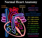
In a normal heart, oxygen-depleted ("blue") blood is pumped from the right side of the heart, through the pulmonary artery, to the lungs where it is oxygenated. The oxygen-rich ("red") blood then returns to the left heart, via the pulmonary veins, and is pumped through the aorta to the rest of the body, including the heart muscle itself.

Transposed vessels can present a large variety of atriovenous, ventriculoarterial and/or arteriovenous discordance. The effects may range from a change in blood pressure to an interruption in circulation, depending on the nature and degree of the misplacement and which vessels are involved.

Terminology

The term "TGV" is often used as a more specific reference to transposition of the great arteries (TGA); however, TGA only relates to the aorta and the pulmonary artery, whereas TGV is a broader term which can relate to these vessels as well as the SVC, IVC, and pulmonary veins.

In it’s strictest sense, transposition of vessels relates only to defects in which two or more vessels have "swapped" positions; in a broader sense, it may be taken to relate to any defect in which a vessel is in an abnormal position.

Variations and similar defects

Simple and complex TGV

In many cases, TGV is accompanied by other heart defects, the most common type being intracardiac shunts such as atrial septal defect (ASD) including patent foramen ovale (PFO), ventricular septal defect (VSD), and patent ductus arteriosus (PDA). Stenosis, or other defects, of valves and/or vessels may also be present.

When no other heart defects are present it is called 'simple' TGV; when other defects are present it is called 'complex' TGV.

Similar defects

The following defects involve abnormal spatial and/or structural arrangement of the great vessels:

  • Total anomalous pulmonary venous connection (TAPVC)
  • Partial anomalous pulmonary Venous Connection (PAPVC)
  • Coarctation of the aorta
  • Cor triatriatum
  • dextro-Transposition of the great arteries (d-TGA)
  • Double outlet right ventricle (DORV)
  • Hypoplastic left heart syndrome (HLHS)
  • levo-Transposition of the great arteries (l-TGA)
  • Overriding aorta
  • Patent ductus arteriosus (PDA)
  • Pulmonary atresia (PA)
  • Unilateral or bilateral Pulmonary arteriovenous malformation (PAVM)
  • Pulmonary sequestration
  • Scimitar syndrome
  • Sinus venosus atrial septal defect (SVASD)
  • Situs inversus
  • Tetralogy of Fallot (TOF)
  • Truncus arteriosus (TA)
  • Vascular rings

Read more at Wikipedia.org


[List your site here Free!]


Double Inlet Left Ventricle with Transposition of the Great Vessels
From Medicine and Health Rhode Island, 6/1/04 by Posteraro, Anthony F III

A 40 year-old male presented to the emergency department with fever and mental status changes. Diagnostic evaluation revealed a brain abscess of uncertain etiology. The patient was known to have a complex uncorrected congenital heart condition, the details of which were incompletely understood.

The patient underwent cardiac MRI examination to further evaluate anatomy and function. Axial black blood Tl weighted images of the chest confirmed dextrocardia (Figure 1) with a single ventricle, situs solitus, and two atrioventricular valves. The patient had transposition of his aorta and pulmonary artery (Figure 2; curved arrow: main pulmonary artery; straight arrow: aorta with a tight pulmonic stenosis demonstrated on cine bright blood images as a large systolic jet (Figure 3) with maximal velocity of 6.8 m/sec. The most accurate anatomic description of this configuration is felt to be double inlet left ventricle with transposition of the great arteries (S,L,L).

A single ventricle is an exceedingly rare cardiac anomaly, estimated to be 3% of all congenital cardiac anomalies.1 By definition a true single ventricle is present when there are two atrioventricular valves emptying into a common ventricle with two great vessels arising from the single ventricle. Most cases are associated with transposition of the great vessels. Since patients have a common mixing chamber for arterial and venous blood, clinical presentation and need for palliation depends on associated lesions.2 Patients such as the one described here, who have balanced lesions with enough pulmonic stenosis to protect the pulmonary circulation, can remain relatively asymptomatic and present later in life with other complications such as peripheral embolization.

REFERENCES

1. Marin-Garcia J, Tandon R, Moller JH, Edwards JE. Single ventricle with transposition. Circulation 1974; 49 : 994.

2. Schwanz SM, Dent CL, Musa NL, Nelson DP. Single-ventricle physiology. Crit Care Clin. 2003; 19: 393-411.

ANTHONY F. POSTERARO III, MD, AND MICHAEL K. ATALAY, MD, PHD

CORRESPONDENCE:

Michael K. Atalay, MD, PhD

e-mail: Matalay@lifespan.org

Anthony F. Posteraro III, MD, is a Resident in the Department of Diagnostic Imaging, Rhode Island Hospital.

Michael K. Atalay, MD, is Director, Cross-sectional Cardiovascular Imaging, in the Department of Diagnostic Inaging, Rhode Island Hospital, and Assistant Professor, Brown Medical School.

Copyright Rhode Island Medical Society Jun 2004
Provided by ProQuest Information and Learning Company. All rights Reserved

Return to Great vessels transposition
Home Contact Resources Exchange Links ebay