Definition
Spinal stenosis is any narrowing of the spinal canal that causes compression of the spinal nerve cord. Spinal stenosis causes pain and may cause loss of some body functions.
Description
Spinal stenosis is a progressive narrowing of the opening in the spinal canal. The spine is a long series of bones called vertebrae. Between each pair of vertebra is a fibrous intervertebral disk. Collectively, the vertebrae and disks are called the backbone. Each vertebra has a hole through it. These holes line up to form the spinal canal. A large bundle of nerves called the spinal cord runs through the spinal canal. This bundle of 31 nerves carries messages between the brain and the various parts of the body. At each vertebra, some smaller nerves branch out from these nerve roots to serve the muscles and tissue in the immediate area. When the spinal canal narrows, nerve roots in the spinal cord are squeezed. Pressure on the nerve roots causes chronic pain and loss of control over some functions because communication with the brain is interrupted. The lower back and legs are most affected by spinal stenosis. The nerve roots that supply the legs are near the bottom of the spinal cord. The pain gets worse after standing for a long time and after some forms of exercise. The posture required by these physical activities increases the stress on the nerve roots. Spinal stenosis usually affects people over 50 years of age. Women have the condition more frequently than men do.
Cervical spinal stenosis is a narrowing of the vertebrae of the neck (cervical vertebrae). The disease and its effects are similar to stenosis in the lower spine. A narrower opening in the cervical vertebrae can also put pressure on arteries entering the spinal column, cutting off the blood supply to the remainder of the spinal cord.
Causes & symptoms
Spinal stenosis causes pain in the buttocks, thigh, and calf and increasing weakness in the legs. The patient may also have difficulty controlling bladder and bowel functions. The pain of spinal stenosis seems more severe when the patient walks downhill. Spinal stenosis can be congenital, acquired, or a combination. Congenital spinal stenosis is a birth defect. Acquired spinal stenosis develops after birth. It is usually a consequence of tissue destruction (degeneration) caused by an infectious disease or a disease in which the immune system attacks the body's own cells (autoimmune disease). The two most common causes of spinal stenosis are birth defect and progressive degeneration of the tissue of the joints (osteoarthritis). Other causes include improper alignment of the vertebrae as in spondylolisthesis, destruction of bone tissue as in Paget's disease, or an overgrowth of bone tissue as in diffuse idiopathic skeletal hyperostosis. The spinal canal is usually more than 11.5 millimeters in diameter. A smaller diameter indicates stenosis. The diameter of the cervical spine ranges is 15-25 millimeters. Any opening under 13 millimeters in diameter is considered evidence of stenosis. Acquired spinal stenosis usually begins with degeneration of the intervertebral disks or the surfaces of the vertebrae or both. In trying to heal this degeneration, the body builds up the spinal column. In the process, the spinal canal can become narrower.
Diagnosis
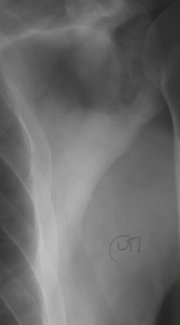
The physician must determine that the symptoms are caused by spinal stenosis. Conditions that can cause similar symptoms include a slipped (herniated) intervertebral disk, spinal tumors, and disorders of the blood flow (circulatory disorders). Spinal stenosis causes back and leg pain. The leg pain is usually worse when the patient is standing or walking. Some forms of spinal stenosis are less painful when the patient is riding an exercise bike because the forward tilt of the body changes the pressure in the spinal column. Doppler scanning can trace the flow of blood to determine whether the pain is caused by circulatory problems. X-ray images, computed tomography scans (CT scans), and magnetic resonance imaging (MRI) scans can reveal any narrowing of the spinal canal. Electromyography, nerve conduction velocity, or evoked potential studies can locate problems in the muscles indicating areas of spinal cord compression.
Treatment
Mild cases of spinal stenosis may be treated with rest, nonsteroidal anti-inflammatory drugs (such as aspirin) and muscle relaxants. Spinal stenosis can be a progressive disease, however, and the source of pressure may have to be surgically removed (surgical decompression) if the patient is losing control over bladder and bowel functions. The surgical procedure removes bone and other tissues that have entered the spinal canal or put pressure on the spinal cord. Two vertebrae may be fused, to eliminate improper alignment, such as that caused by spondylolisthesis. For surgery, patients lie on their sides or in a modified kneeling position. This position reduces bleeding and places the spine in proper alignment. Alignment is especially important if vertebrae are to be fused. Surgical decompression can eliminate leg pain and restore control of the legs, bladder, and bowels, but usually does not eliminate lower back pain. Physical therapy and massage can help reduce the symptoms of spinal stenosis. An exercise program should be developed to increase flexibility and mobility. A brace or corset may be worn to improve posture. Activities that place stress on the lower back muscles should be avoided.
Prognosis
Surgical decompression does not stop the degenerative processes that cause spinal stenosis, and the condition can develop again. Nevertheless, most patients achieve good results with surgical decompression. The patient will probably continue to have lower back pain after the surgical procedure.
Key Terms
- Computed tomography (CT)
- An imaging technique in which cross-sectional x rays of the body are compiled to create a three-dimensional image of the body's internal structures.
- Congenital
- Present before birth. The term is used to describe disorders that developed in the fetal stage.
- Doppler scanning
- A procedure in which ultrasound images are used to watch a moving structure such as the flow of blood or the beating of the heart.
- Electromyography
- A test that uses electrodes to record the electrical activity of muscle. The information gathered is used to find disorders of the nerves that serve the muscles.
- Evoked potential
- A test of nerve response that uses electrodes placed on the scalp to measure brain reaction to a stimulus such as a touch.
- Magnetic resonance imaging (MRI)
- An imaging technique that uses a large circular magnet and radio waves to generate signals from atoms in the body. These signals are used to construct images of internal structures.
- Nerve conduction velocity test
- A test that measures the time it takes a nerve impulse to travel a specific distance over the nerve after electronic stimulation.
- Stenosis
- The narrowing or constriction of a channel or opening.
Further Reading
For Your Information
Books
- Berkow, Robert, ed. Merck Manual of Medical Information. Whitehouse Station, NJ: Merck Research Laboratories, 1997.
- Dee, Roger, et al. Principles of Orthopaedic Practice. New York: McGraw-Hill Health Professional Books, 1997.
- Larsen, D.E., ed. Mayo Clinic Family Health Book: New York. William Morrow and Company, Inc., 1996.
Gale Encyclopedia of Medicine. Gale Research, 1999.


