Definition
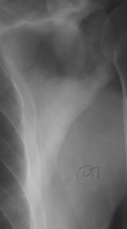
Skull x rays are performed to examine the nose, sinuses, and facial bones. These studies may also be referred to as sinus x rays. X-ray studies produce films, also known as radiographs, by aiming x rays at soft bones and tissues of the body. X-ray beams are similar to light waves, except their shorter wavelength allows them to penetrate dense substances, producing images and shadows on film.
Purpose
Doctors may order skull x rays to aid in the diagnosis of a variety of diseases or injuries.
Sinusitis
Sinus x rays may be ordered to confirm a diagnosis of sinusitis, or sinus infection.
Fractures
A skull x ray may detect bone fractures resulting from injury or disease. The skull x ray should clearly show the skull cap, jaw bones, and facial bones.
Tumors
Skull radiographs may indicate tumors in facial bones, tissues, or the sinuses. Tumors may be benign (not cancerous) or malignant (cancerous).
Other
Birth defects (referred to as congenital anomalies) may be detected on a skull x ray by changes in bone structure. Abnormal tissues or glands resulting from various conditions or diseases may also be shown on a skull radiograph.
Precautions
As with any x-ray procedure, women who may be pregnant are advised against having a skull x ray if it is not absolutely necessary. However, a lead apron may be worn across the abdomen during the procedure to protect the fetus. Children are also more sensitive to x-ray exposure. Children of both sexes should wear a protective covering (a lead apron) in the genital/reproductive area. In general, skull x-ray exposure is minimal and x-ray equipment and procedures are monitored to ensure radiation safety.
Description
Skull or sinus x rays may be performed in a doctor's office that has x-ray equipment and a technologist available. The exam may also be performed in an outpatient radiology facility or a hospital radiology department.
In many instances, particularly for sinus views, the patient will sit upright in a chair, perhaps with the head held stable by a foam vise. A film cassette is located behind the patient. The x-ray tube is in front of the patient and may be moved to allow for different positions and views. A patient may also be asked to move his or her head at various angles and positions.
In some cases, technologists will ask the patient to lie on a table and will place the head and neck at various angles. In routine skull x rays, as many as five different views may be taken to allow a clear picture of various bones and tissues. The length of the test will vary depending on the number of views taken, but in general, it should last about 10 minutes. The technologist will usually ask a patient to wait while the films are being developed to ensure that they are clear before going to the radiologist.
Preparation
There is no preparation for the patient prior to arriving at the radiology facility. Patients will be asked to remove jewelry, dentures, or other metal objects that may produce artifacts on the film. The referring doctor or x-ray technologist can answer any questions regarding the procedure. Any woman who is, or may be, pregnant should tell the technologist.
Aftercare
There is no aftercare required following skull or sinus x-ray procedures.
Risks
There are no common side effects from skull or sinus x ray. The patient may feel some discomfort in the positioning of the head and neck, but will have no complications. Any x-ray procedure carries minimal radiation risk, and children and pregnant women should be protected from radiation exposure to the abdominal or genital areas.
Normal results
Normal results should indicate sinuses, bones, tissues, and other observed areas are of normal size, shape, and thickness for the patient's age and medical history. Results, whether normal or abnormal, will be provided to the referring doctor in a written report.
Abnormal results
Abnormal results may include:
Sinusitis
Air in sinuses will show up on a radiograph as black, but fluid will be cloudy or white (opaque). This helps the radiologist to identify trapped fluids in the sinuses. In chronic sinusitis, the radiologist may also note thickening or hardening of the bony wall of an infected sinus.
Fractures
Radiologists may recognize even tiny facial bone fractures as a line of defect.
Tumors
Tumors may be visible if the bony sinus wall is distorted or destroyed. Abnormal findings may result in follow-up imaging studies.
Other
Skull x rays may also detect disorders that show up as changes in bone structure, such as Paget's disease of the bone or acromegaly (a disorder associated with excess growth hormones from the pituitary gland). Areas of calcification, or gathering of calcium deposits, may indicate a condition such as an infection of bone or bone marrow (osteomyelitis).
Key Terms
- Radiograph
- The actual picture or film produced by an x-ray study.
- X ray
- A form of electromagnetic radiation with shorter wavelengths than normal light. X rays can penetrate most structures.
Further Reading
For Your Information
Books
- Illustrated Guide to Diagnostic Tests, edited by D. Weinstock, et al. Springhouse, PA: Springhouse Corporation, 1998.
Organizations
- Cancer Information Clearinghouse, National Cancer Institute. Building 31, Room 10A24, 9000 Rockville Pike, Bethesda, MD 20892. (800) 4-Cancer. http://www.nci.nih.gov.
- The National Head Injury Foundation, Inc. 1140 Connecticut Ave. NW, Suite 812, Washington, DC 20036. (800) 444-NHIF.
- Radiological Society of North America. 2021 Spring Rd., Suite 600, Oak Brook, IL 60521-1860. (708) 571-2670. http://www.rsna.org.
Gale Encyclopedia of Medicine. Gale Research, 1999.


