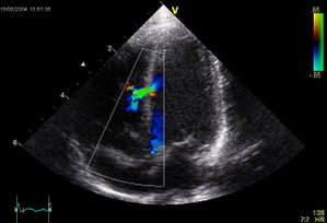
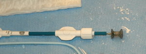
Echocardiographic image of a moderate ventricular septal defect in the mid-muscular part of the septum. The size and location is typical for a VSD in a new-born child.  The trace in the lower left shows the flow during one complete cardiac cycle and the red mark the time in the cardiac cycle that the image was captured. Colours are used to represent the velocity of the blood.  Flow is from the left ventricle (right on image) to the right ventricle (left on image).A nitinol device for closing muscular VSDs, 4 mm diameter in the centre. It is shown mounted on the catheter into which it will be withrawn during insertion.
Find information on thousands of medical conditions and prescription drugs.

Ventricular septal defect

A ventricular septal defect (or VSD) is a defect in the ventricular septum (the wall dividing the left and right ventricles of the heart). more...

Home
Diseases
A
B
C
D
E
F
G
H
I
J
K
L
M
N
O
P
Q
R
S
T
U
V
Vaccinophobia
VACTERL association
Vaginismus
Van der Woude syndrome
Van Goethem syndrome
Varicella Zoster
Variegate porphyria
Vasculitis
Vasovagal syncope
VATER association
Velocardiofacial syndrome
Ventricular septal defect
Vipoma
Viral hemorrhagic fever
Vitamin B12 Deficiency
Vitiligo
VLCAD deficiency
Von Gierke disease
Von Hippel-Lindau disease
Von Recklinghausen disease
Von Willebrand disease
Vulvodynia
W
X
Y
Z
Medicines

The ventricular septum consists of a muscular (inferior) and membranous portion (superior). The membranous portion (which is close to the atrioventricular node) is most commonly affected.

Congential VSDs are collectively the most common congenital heart defect.

Diagnosis

VSDs can be detected by cardiac auscultation, as they typically cause systolic murmurs. Confirmation of findings from cardiac auscultation can be obtained with a cardiac ultrasound (echocardiography) (less invasive) and cardiac catheterization (more invasive).

Auscultation is generally considered sufficient for ruling-out a significant VSD, if done by a pediatric cardiologist. This holds true as long as the pressures on the right side of the heart is low.

Pathophysiology

Large VSDs result in a significant left-to-right shunt and increase load on the right ventricle. If untreated, they result in hypertrophy of the right ventricle, which ultimately leads to right heart failure and death.

Treatment

Treatment is either surgical (open or percutaneous endovascular) or conservative. Smaller congenital VSDs often close on their own (as the heart grows) and are thus treated conservatively. Open surgical procedures require a heart-lung machine and are done with a median sternotomy. Percutaneous endovascular procedures are less invasive and can be done on a beating heart, but are only suitable for certain patients. Repair of most VSDs is complicated by the fact that the conducting system of the heart is in the immediate vicinity.

Epidemiology

VSDs are the most common congenital cardiac anomaly. It is found in 30% of all newborns with a congenital heart defect, or about 2-3 per 1000 births.

Congential VSDs are frequently associated with other congential conditions, such as Down syndrome.

A VSD can form a few days after a myocardial infarction (heart attack) due to mechanical tearing of the septal wall, before scar tissue forms, when macrophages start remodeling the dead (heart) tissue.

Read more at Wikipedia.org


[List your site here Free!]


Interventricular septal flattening observed on the myocardial perfusion images strongly correlate with right ventricular overload
From CHEST, 10/1/05 by Mohammad-Reza Movahed

PURPOSE: Background: Flattening of the interventricular septum (D-shaped Septum) detected during echocardiographic examination is correlated with significant right ventricular (RV) overload. There are no reports of this finding with cardiac gated SPECT imaging. We report an observational study correlating this finding with the presence-of RV overload.

METHODS: Method: Retrospectively we compared eight cases with flattening of the interventricular septum on cardiac gated SPECT imaging of which echocardiographic correlations and clinical data were available for the presence of RV overload.

RESULTS: Results: All patients but one had pulmonary hypertension ranging from 42 to 52 mmHg measured by echocardiographic doppler studies. Except one all patients had reasons for RV overload (chronic obstructive pulmonary disease in three, history of atrial septal defect in three, pulmonary embolism in one and obstructive sleep apnea in one). Septal flattening present on gated SPECT images was seen in 50% of the cases by echocardiography. Other signs of RV overload (RV enlargement, RV hypertrophy) were observed by echocardiography in five patients and by the gated SPECT in seven patients.

CONCLUSION: Conclusion: The presence of interventricular septal flattening on gated SPECT studies correlates with right ventricular overload and should be routinely assessed during interpretation of gated SPECT studies.

CLINICAL IMPLICATIONS: We suggest that more attention should be given to the shape of the interventricular septum during interpretation of gated SPECT studies and the term of septal flattening should be routinely used and reported similar to echocardiographic descriptions of the septum in patients with suspected RV overload.

DISCLOSURE: Mohammad-Reza Movahed, None.

Mohammad-Reza Movahed MD * Absalam Hepner MD Paul Lazotte DO Norah Milne MD University of California, Irvine Medical Center, Orange, CA

COPYRIGHT 2005 American College of Chest Physicians
COPYRIGHT 2005 Gale Group

Return to Ventricular septal defect
Home Contact Resources Exchange Links ebay