Abstract
Cowden's disease is a hereditary disorder characterized by oropharyngeal fibrosis and multiple hamartomas with potential malignant changes. We treated a 47-year-old man who had fibrotic lesions on the left vocal fold and an extensive amount of papillomatous lesions on the mucosa of the lips, tongue, and pharyngeal wall and on the skin of the axillae and buttocks. The pattern of distribution and the histopathologic features of these mucocutaneous lesions were diagnostic of Cowden's disease. To the best of our knowledge, this is the first reported case of Cowden's disease involving a vocal fold.
Introduction
Cowden's disease (multiple hamartoma syndrome), first described in 1963 by Lloyd and Dennis, is a rare autosomal-dominant disease. [1-4] Its features usually include facial tricholemmomas, acral and palmoplantar keratoses, multiple oral papules, and mucosal fibrosis with a cobblestone appearance. Cowden's disease is also associated with multiple hamartomas of the gastrointestinal tract and cancers of the breast, thyroid, uterus, ovary, lung, skin, and gastrointestinal tract. [3,5] Its close association with Lhermitte-Duclos disease, a similar disorder characterized by cerebellar hypertrophy, suggests that these fibrotic lesions represent the continuum of a spectrum. [6,7]
To our knowledge, there has been no previous report in the literature of Cowden's disease in which the fibrotic lesions affected a vocal fold. In this article, we describe one such case to illustrate that the pathology of Cowden' s disease can be manifested in the larynx. We also discuss the differential diagnosis of multiple mucocutaneous fibrosis.
Case report
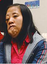
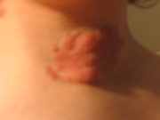
A 47-year-old man came to the ENT department for investigation of hoarseness, which he had had since childhood. Oral examination revealed a diffuse induration over the lower lip and multiple papules and nodules on the tongue (figure 1). The posterior pharyngeal wall had a cobblestone appearance. A 2-cm submental lymph node was identified on palpation. In addition, there were papillomatous lesions with pigmentation on the axillae (figure 2) and buttocks. Flexible laryngoscopy revealed that the left vocal fold was thickened and had an irregular free edge (figure 3). Subsequent examination under general anesthesia revealed the presence of induration on the posterior pharyngeal wall and the base of the tongue.
Findings on ultrasonography of both the thyroid and abdomen were normal. Chest x-ray showed no features of mediastinal fibrosis. Sputum culture did not detect any bacteria or acid-fast bacilli. Autoantibody titers were normal, and tests for anti-DNA and anti-extractable nuclear antigens were negative. The erythrocyte sediment rate was 11 mm/hour. The patient's serum profile was not characteristic or suggestive of connective tissue disease. Analysis of fine-needle aspiration of the submental lymph node showed that it was of a reactive nature. During laryngoscopy, biopsies were obtained from the indurated laryngeal wall, the thickened vocal fold, and the lips. All these samples exhibited hyperplasia of the squamous epithelium with acanthosis, parakeratosis, and hyperkeratosis. Analysis of the squamous cells revealed normal maturation without dysplasia or malignancy (figure 4). The underlying stroma featured dense fibrosis and a nonspecific lymphocytic infiltrate. There was no evidence of amyloid deposits, granulomatous inflammation, or neoplasia.
Subsequently, the biopsy samples taken from the skin of the axillae and buttocks were also submitted for histo-pathologic examination. Acanthosis, papillomatosis, hyperkeratosis of the epidermis, and elongation of the rete ridges were evident, lathe dermis, thick bands of collagen were also noted. The histologic findings were those of keratotic papules. In view of the constellation of clinical features--oropharyngeal fibrosis and papillary lesions on the pharynx, larynx, lips, left vocal fold, and skin-a diagnosis of Cowden's disease was made.
Discussion
The coexistence of multiple mucocutaneous and laryngeal fibroses poses a diagnostic challenge to both the clinician and the pathologist. Making the definitive diagnosis depends on recognizing the pattern of the clinical features and on the histology of the corresponding lesions. In our patient, the lesions of the lip, left vocal fold, axillae, and buttocks demonstrated the characteristic features of hyperkeratosis and nodular proliferation of bands of fibrous tissue. These findings confirmed the diagnosis of Cowden's disease. The presence of fibrotic lesions on the vocal fold makes this case unique.
In addition to Cowden's disease, the differential diagnosis includes neurofibromatosis, amyloidosis, and multifocal idiopathic fibrosis:
Neurofibromatosis. Neurofibromatosis (von Recklinghausen's disease) is characterized by multiple subcutaneous nodules and cafe au lait spots. It is known to affect multiple organs, including the larynx. [8,9] The laryngeal lesions are predominantly limited to the aryepiglottic folds, false vocal folds, and the arytenoids. In our patient, the absence of neurofibromas and schwannomas in the excised tissues made a diagnosis of neurofibromatosis unlikely.
Amyloidosis. Localized involvement of the larynx by amyloidosis has been reported to affect the vocal folds, false folds, and ventricles. [10] Gottschalk et al reported a single case of combined laryngeal and dermal amyloidosis in which the cutaneous lesions were small and macular. [11] In our patient, no evidence of amyloid deposits was noted, either morphologically or on Congo red staining.
Multifocal idiopathic fibrosis. In patients with Cowden's syndrome, retroperitoneal fibrosis, mediastinal fibrosis, sclerosing cholangitis, Riedel's thyroiditis, and inflammatory pseudotumor of the orbit can all manifest, either alone or in combination. These inflammatory fibrosclerotic lesions characteristically exhibit a florid proliferation of fibroblasts on a cellular background of mixed inflammatory infiltrates of lymphocytes, plasma cells, and eosinophils. [12] The end-stage lesions are densely sclerotic, and they can compress and distort adjacent normal structures. Involvement of the larynx is extremely rare, and only one case has been reported to involve the upper aerodigestive tract; in the latter case, a dense fibrosis was noted in the floor of the mouth, the tongue, the pharynx, and the larynx, but not in the vocal folds. [13] In our patient, the absence of prominent fibroblastic proliferation and inflammatory-cell infiltrate in addition to the normal findings on abdominal, mediastinal, and thorac ic imaging led us to rule out this possibility.
Early diagnosis of Cowden's disease is critical, and lifelong followup is necessary because associated malignancies might take decades to achieve their full clinical manifestation. Because Cowden's disease is associated with carcinomas of the breast, thyroid, uterus, ovary, lung, skin, and gastrointestinal tract, a thorough initial systemic workup and close long-term observation of all these regions is indicated. [3,5]
References
(1.) Lloyd KM, Dennis M. Cowden's disease: A possible new symptom complex with multiple systemic involvement. Ann Intern Med 1963;58:l36-42.
(2.) Weary PE, Gorlin RJ, Gentry WC, Jr., et al. Multiple hamartoma syndrome (Cowden's disease). Arch Dermatol 1972;106:682-90.
(3.) Starink TM, van der Veen JP, Arwert F, et a!. The Cowden syndrome: A clinical and genetic study in 21 patients. Clin Genet 1986;29:222-33.
(4.) Thyresson HN, Doyle JA. Cowden's disease (multiple hamartoma syndrome). Mayo Clin Proc 1981;56:179-84.
(5.) Mallory SB. Cowden syndrome (multiple hamartoma syndrome), Dermatol Clin 1995;13:27-31.
(6.) Chapman MS, Perry AE, Baughman RD. Cowden's syndrome, Lhermitte-Duclos disease, and sclerotic fibroma. Am J Dermatopathol 1998;20:413-6.
(7.) Albrecht S, Haber RM, Goodman JC, Duvic M. Cowden syndrome and Lhermitse-Duclos disease. Cancer 1992;70:869-76.
(8.) Chang-Lo M. Laryngeal involvement in Von Recklinghausen's disease: A case report and review of the literature, Laryngoscope 1977;87:435-42.
(9.) O'Connor AF, Freeland AP. Neonatal laryngeal neurofibromatosis. Ear Nose Throat J 1980;59:174-7.
(10.) Barnes EL, Jr., Zafar T. Laryngeal amyloidosis: Clinicopathologic study of seven cases. Ann Otol Rhinol Laryngot 1977:86:856-63.
(11.) Gottschalk HR, Graham JH, Kohut RI. Macular amyloidosis with localized amyloidosis of upper air passages. Arch Dermatol 1975;lll:1017-9.
(12.) Comings DE, Skubi KB, Van Eyes J, Motulsky AG. Familial moltifocal fibrosclerosis. Findings suggesting that retroperitoneal fibrosis, mediastinal fibrosis, sclerosing cholangitis, Riedel's thyroiditis, and pseudotumor of the orbit may be different manifestations of single disease. Ann Intern Med 1967:66:884-92.
(13.) Violaris NS, Windle-Taylor PC. Idiopathic fibrosis of the upper aero-digestive tract. J Laryngol Otol 1989:103:333-4.
COPYRIGHT 2001 Medquest Communications, Inc.
COPYRIGHT 2001 Gale Group


