ABSTRACT
SHARMA, S.P, AMANFU, W & LOSHO, TC. 2000. Bovine borreliosis in Botswana. Onderstepoort Journal of Veterinary Research, 67:221-223
Clinical Borrelia theileri infection was reported for the first time in cattle from Botswana concurrent with Babesia bovis and Theileria mutans infections. Two animals, an ox and a cow of the Tswana breed demonstrated clinical signs of fever, haemoglobinuria, inappetance, diarrhoea, pallor of mucous membranes, enlarged superficial lymph nodes and rough hair coats. Examination of the blood smears from the affected animals revealed numerous B. theileri, and very few B. bovis and T mutans organisms. Oxytetracycline was administered parenterally to all the animals in the herd.The ox, being extremely weak and recumbent for the previous 4-5 days, succumbed to death the day after the examination. The clearance of spirochaetes from the blood circulation and recovery of the cow three days after treatment with oxytetracycline suggest an involvement of B. theileri in producing clinical disease.
Keywords: Babesia bovis, Borrelia theileri, cattle blood, Theileria mutans
INTRODUCTION
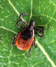
Bovine spirochaetosis caused by Borrelia burgdorferi and Borrelia theileri has been reported from several countries spanning almost every continent of the world. Lyme disease caused by B. burgdorferi, one of the fast emerging zoonoses, is transmitted by Ixodes spp. of ticks parasitizing small wild animals which are considered to be reservoirs of this spirochaetal organism which also affects cattle, sheep, horses, dogs and humans. Borreliosis caused by B. theileri has been reported from cattle, sheep and horses from several countries in Africa, South America, Europe and Australia. Several tick species, Rhipicephalus evertsi evertsi, Boophilus decoloratus, Boophilus australis, Boophilus microplus and Boophilus annulatus are known to be vectors of B. theileri which cause a mild infection and is often associated with babesiosis (Bishop 1994). The present report describes for the first time, a natural B. theileri infection in an ox and a cow, in Botswana.
Two adult cattle, an ox and a cow of the Tswana breed from Molepolole-Kweneng district in a herd of 23 cattle were reported to be suffering from fever, haemoglobinuria, inappetance and diarrhoea for the previous 4-5 days. The sick and in-contact animals were examined physically and blood smears, whole blood, faecal and urine samples were collected for investigation at the National Veterinary Laboratory (NVL), Gaborone. Ticks were also collected from the diseased animals for identification. Oxytetracycline (Terramycin-Pfizer) was administered intramuscularly to all 23 animals in the herd at the dose of 15 mg/ kg body mass as a possible prophylactic measure. On physical examination, the affected animals appeared dull and depressed with poor bodily conditions. Conjunctival and other visible mucous membranes were pale and the prescapular and parotid lymph nodes were enlarged. The hair coats of both animals were rough and lusterless.The body temperatu res of the ox and cow were 39,8 'C and 39,5 deg C, respectively. The ox, which was recumbent and extremely weak when examined, died the day after the examination and collection of samples.
Examination of Giemsa-stained blood smears from the ox demonstrated a mixed haemoparasitic infection consisting of numerous spiral and knotted organisms indistinguishable from B. theileri, and very few Babesia bovis and Theileria mutans parasites (Fig. 1). The blood sample of the cow showed the presence of a mild parasitaemia consisting of few B. theileri, B. bovis and T mutans organisms. All spirochaetal organisms examined were within the range of 918,5 Ij in length, with the average being 13,2 p. No haemoparasite could be observed in any of the incontact animals, except three cows that showed a few T mutans organisms in blood smears. Urinalysis of the diseased animals revealed the presence of albumin, bile salts and erythrocytes. Anaemia, indicated by prominent reticulocytosis, poikilocytosis and anisocytosis, was evident in the blood smears of both affected animals.
Differential leukocytic counts revealed monocytosis in both animals. Haematological values are presented in Table 1. In the faecal sample of the ox, 300 Trichostrongylus spp. egg/g were demonstrated, whereas that of the cow was negative for helminthic eggs and coccidia.
Leptospira organisms could not be detected in the urine by dark field microscopy. Two guinea pigs inoculated intraperitoneally with the ox blood subsequently proved negative for leptospira organisms. Sera were negative for leptospiral antibodies using the ELISA technique. Serum calcium levels of the ox and the cow were 3,0 and 2,7 mmol/p and those of phosphorus were 2,5 and 2,0 mmol/V, respectively. Ticks collected from the cattle were all identified as Rhipicephalus everts! evertsi.
The spirochaetal organisms observed in the blood samples from the ox and cow were morphologically similar to those described by Callow 1967, Van Heerden & Reyers 1984 and Bishop 1994.
Bovine borreliosis has been reported in cattle of South Africa (Neitz 1956), Nigeria (Trees 1978), Ghana (Assoku 1979), Australia (Callow & Hoyte 1961; Callow 1967), Mauritius (Barre & Morel 1983), USA (Smith, Miranpuri, Adams & Ahrens 1985), Democratic Republic of Congo (Matton & Van Melckebeke 1990), Sweden (Hovamark, Asbrink, Schwan, Henderstedt & Christensson 1986) and in several other countries. However, this report constitutes the first of its kind in Botswana. It is difficult to state with certainty whether the clinical manifestations of fever, haemoglobinuria, diarrhoea, pallor of mucous membranes and enlargement of lymph nodes in these two animals were caused by borreliosis alone, or in conjunction with babesiosis. Though B. theiled is considered to be mildly pathogenic, the clinical syndrome comprising fever, anaemia and haemoglobinuria resembling babesiosis has been reported in some cases of borreliosis (Callow 1967). The disappearance of spirochaetes from the blood circulation and complete recovery of the cow three days after treatment with oxytetracycline may suggest an involvement of B. theileri in producing the clinical manifestations observed. R. evertsi evertsi ticks that were collected from affected animals have been incriminated as vectors of borreliosis (Bishop 1994).
ACKNOWLEDGEMENTS
The authors thank Dr M.V. Raborokgwe, Director, Department of Animal Health and Production, Botswana for granting permission to publish this research article, and Dr J.F.C. Nyange, Dr M.G. Binta and Mr B.K. Reasentse of National Veterinary Laboratory (NVL) for their help and suggestions.
REFERENCES
ASSOKU, R.K.G. 1979. A study of the incidence of blood borne parasites of livestock in Southern Ghana. Bulletin of Animal Health and Production in Africa, 27:29-39.
BARRE, N. & MOREL, PC. 1983. Ticks (Africana, Ixodoidea) of the Mascarene Islands and tick-borne disease. Revue d'Elevage et de Medecine Veterinare des Pays Tropicaux, 36:371377.
BISHOP, G.C. 1994. Borrelia theileri infection, in Infectious diseases of livestock with special reference to Southern Africa, edited by J.A.W. Coetzer, G.R. Thomson & R.C. Tustin. Oxford: Oxford University Press.
CALLOW, L.L. & HOYTE, H.M.D. 1961. Transmission experiments using Babesia bigemina, Theileria mutans, Borrelia sp. And cattle tick, Boophilus microplus. Australian Veterinary Journal, 37:381-390.
CALLOW, L.L. 1967. Observation on tick-transmitted spirochaetes of cattle in Australia and South Africa. British Veterinary Journal, 123:492-496.
HOVAMARK, A., ASBRINK, E., SCHWAN, 0., HENDERSTEDT, B. & CHRISTENSSON, D. 1986. Antibodies to Borrelia spirochaetes in sera from Swedish cattle and sheep. Acta Merinaria Scandinavica, 27:479-485.
MATTON, P. & VAN MELCKEBEKE, H. 1990. Bovine borreliosis: Comparison of simple methods for detection of spirochaetes in blood. Tropical Animal Health and Production, 22:147-152.
SMITH, R.D., MIRANPURI, G.S., ADAMS, J.H. & AHRENS, E.H. 1985. Borrelia theileri: Isolation from ticks (Boophilus microplus) and tick-borne transmission between splenectomized calves. American Journal of Veterinary Research, 46:13961398.
TREES, A.J. 1978. The transmission of Borrelia theileri by Boophilus annulatus. Tropical Animal Health and Production, 10: 93-94.
VAN HEERDEN, J. & REYERS, F. 1984. Borrelia sp. infection in a horse. Journal of the South African Veterinary Association, 55:41-43.
S.P SHARMA, W. AMANFU and TC. LOSHO
National Veterinary Laboratory, Private Bag 0035, Gaborone, Botswana
Accepted for publication 22 May 2000-Editor
Copyright Onderstepoort Veterinary Institute Sep 2000
Provided by ProQuest Information and Learning Company. All rights Reserved


 Home
Home A
A Babesiosis