A 69-year-old woman diagnosed as having Charcot-Marie-Tooth disease (CMT) for 30 years' duration died of respiratory failure secondary to diaphragmatic weakness. At autopsy, identical neuropathic changes were found in phrenic and other affected somatic peripheral nerves. We conclude that the phrenic nerve may be involved by the neuropathy of at least some forms of CMT.
Charcot-Marie-Tooth disease (CMT) is a collection of chronic degenerative neuropathic conditions with differing clinical and pathologic manifestations and modes of heritability. It has previously been thought that the disease spares the phrenic nerves. We present a case of CMT of 30 years' duration with respiratory dysfunction secondary to diaphragmatic weakness. Phrenic neuropathic changes identical to the other affected somatic peripheral nerves together with diaphragmatic neurogenic atrophy were identified postmortem.
CASE REPORT
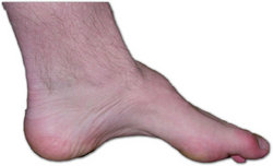
A 69-year-old woman had been diagnosed as having CMT 30 years previously. Her neurologic difficulty began at age 31, when she noted weakness of her feet and ankles and the development of bilateral footdrop. Approximately three years later, she noted wasting and weakness in the small muscles of her hands, sensory loss of both feet associated with an unsteady gait, and falling occasionally. No hypertrophic changes were noted in her peripheral nerves.
Peripheral nerve conduction study of both upper and lower extremities that were done 20 years ago showed reduced motor nerve conduction velocity; right lateral popliteal nerve at 20 m/s, right ulnar nerve at 29 m/s (normal >45 m/s). Similarly, sensory nerve conduction velocity was also delayed in the right ulnar nerve at 44 m/s (normal>55 m/s). The striking feature was a very low amplitude of the sensory nerve action potential at 2 mV (normal 15 to 20 mV).
In the ensuing years, she suffered extensive distal weakness together with loss of sensation in both feet and hands with painful dysesthesia. The patient had experienced difficulty with breathing in a supine position for several years and had adapted by sleeping on an incline on her side. Eight months prior to her death, she noted more difficulty with her breathing and increased weakness and tingling of her hands.
The patient was an exsmoker but had been a nonsmoker for 40 years. She had no known lung disease. Chest radiographs, other than showing reduced lung volumes, were normal. She had suffered myocardial infarctions in 1977 and 1985 with residual stable exertional angina, but no evidence of ongoing congestive heart failure. She had hypertension which was well controlled. The patient's father and brother had similar difficulty with their extremities. She had two children with no evidence of impairment on nerve conduction velocity and EMG studies.
On examination two months prior to her death, the patient appeared frail, undernourished, and emotionally labile. Pertinent physical findings included poor thoracic expansion and thoracoabdominal paradox in the semirecumbent position. Breath sounds were decreased; there were no adventitious sounds. Cardiac sounds were distant but normal. The jugular venous pressure was 2 cm above the sternal angle, and no peripheral edema was present. Neurologic examination results showed dysphonia and dysarthria and mild bilateral weakness and wasting of facial muscles. There was weakness and wasting of distal upper musculature with fasciculations. Marked atrophy of both proximal and distal muscles of the lower limbs with bilateral footdrop was noted. Deep tendon reflexes were absent. There was marked loss of vibration and position sense in a glove-and-stocking distribution to knees and elbows.
The patient died of bronchopneumonia before diaphragmatic studies could be done. Extensive neuropathologic examination at autopsy revealed peripheral neuropathy in both the sensory and motor nerves, most marked in phrenic nerve and in lower limbs distally with neuronal loss from: (a) dorsal root ganglia with pallor of dorsal columns, (b) anterior horns, spinal cord. Also, severe neurogenic atrophy, secondary to peripheral neuropathy, was noted affecting the diaphragm, to a moderate degree (Fig 1), and the limbs, bilaterally, most marked distally, chiefly in the lower limbs. There was evidence of old hemorrhage and thrombosis in the pons. Specific examination of the phrenic nerve close to its insertion in the diaphragm revealed few surviving myelinated axons with severe decrease of large but not of small fibers on neurofilament and myelin (Fig 2) stains. These changes were identical to those found in median, sciatic, and sural nerves. In contrast, the vagus nerve was largely unaffected. No hypertrophic, specifically "onion bulb," changes were noted in any nerve. Examination showed no other CNS lesion.
DISCUSSION
According to the classification proposed by Dyck et al,[1-3] CMT disease includes cases of hereditary motor neuropathy (HMN) and hereditary motor and sensory neuropathy (HMSN), the common feature of which is the slowly progressive and predominantly distal and lower limb effects of the disease process.
The first case of diaphragmatic dysfunction described in two siblings with CMT by Chan et al[4] was published in 1987. A letter[5] in response to this article outlined another patient with CMT and respiratory failure. More recently, Laroche et al[6] reported two CMT patients with marked diaphragmatic weakness, and respiratory muscle dysfunction was documented in a clinic-based cohort of ten patients with HMSN type 1.[7] We described a 69-year-old patient with a 30-year history of CMT with symptomatic respiratory dysfunction thought clinically to be due to diaphragmatic weakness. At autopsy, changes in the phrenic nerve were identical to those in the affected somatic nerves. Additionally, neurogenic atrophy of the diaphragm mirrored that in distal musculature. The clinical presentation, nerve conduction velocity, and EMG studies of this patient are consistent with HMSN type 2. The neuropathologic documentation in this case demonstrated that phrenic nerve, which is part of the peripheral nervous system, can be substantially involved in certain patients with advanced CMT.
CMT disease, being a peripheral neuropathic process, tends to predominantly affect somatic peripheral nerves with long axons, eg, popliteal or sural. But like any generalized degenerative neuropathic disease, in advanced cases peripheral nerves with shorter axons such as the phrenic may also be affected. We submit that diaphragmatic weakness due to phrenic neuropathy be considered in any patient with CMT who complains of respiratory difficulty.[8] It remains to be ascertained whether phrenic nerve involvement is a common long-term complication of CMT, whether it occurs in all types of CMT, and whether it follows the same conservative hereditary pattern as other markers of the disease.
REFERENCES
[1] Harding AE. Inherited neuronal atrophy and degeneration predominantly of lower motor neurons. In: Dyck PJ, Thomas PK, Lambert EH, Bange R, eds. Peripheral neuropathy, 2nd ed. Philadelphia: WB Saunders Co, 1984:1537-56
[2] Dyck PJ, Lambert EH. Lower motor and primary sensory neuron diseases with peroneal muscular atrophy: I. Neurologic, genetic and electrophysiologic findings in hereditary polyneuropathies. Arch Neurol 1968; 18:603-18
[3] Dyck JP, Lambert EH. Lower motor and primary sensory neuron diseases with peroneal muscular atrophy: I. Neurologic, genetic and electrophysiologic findings in various neuronal degeneration. Arch Neurol 1968; 18:619-25
[4] Chan CK, Mohsenin V, Loke J, Virgulto J, Sipski ML, Ferranti R. Diaphragmatic dysfunction in siblings with hereditary motor and sensory neuropathy (Charcot-Marie-Tooth disease). Chest 1987;91:567-70
[5] Dyer EL, Callahan AS. Charcot-Marie-Tooth disease and respiratory failure (letter). Chest 1987; 92:957
[6] Laroche CM, Carroll N, Moxham J, Stanley NN, Courtenay Evans RJ, Green M. Diaphragm weakness in Charcot-Marie-Tooth disease. Thorax 1988; 43:478-79
[7] Eichacker PQ, Spiro A, Sherman M, Lazar E, Reichel J, Dodick F. Respiratory muscle dysfunction in hereditary motor sensory neuropathy, type I. Arch Intern Med 1988; 148:1759-40
[8] Schmalstieg EJ, Peters BH, Schochet SS, Findlay SR. Neuropathy presenting as prolonged dyspnea. Arch Neurol 1977; 34:473-76
COPYRIGHT 1989 American College of Chest Physicians
COPYRIGHT 2004 Gale Group


