Case report
We describe the clinical features of calcifying tendonitis Win the medial head of gastrocnemius in three elderly female patients. The presenting symptom was chronic pain in the posteromedial area of the knee in two patients and acute pain in the back of the knee in one. All had limitation of movement of the knee and marked tenderness in the region of the tendinous origin of the medial head of gastrocnemius with posterior knee pain induced by stretching the tendon. An injection of 1% lidocaine and steroid into the tendon resulted in temporary relief from pain and improved movement.
J Bone Joint Surg [Br] 2002;84-B:431-2.
Received 8 December 2000; Accepted after revision 28 June 2001
In 1981, Martel et al1 first reported the characteristic radiological appearance of calcification in the tendon of gastrocnemius associated with the deposition of crystals of calcium pyrophosphate dehydrate (CPPD). Although a high incidence of calcification within the tendinous origin of the medial head of gastrocnemius has been described,2,3 associated tendonitis has not been reported.
We present the clinical features of three patients with calcifying tendonitis of gastrocnemius.
Case reports
Case 1. A 67-year-old woman farmer presented in March 1998 with a one-year history of posteromedial pain in the left knee. There was a tender swelling measuring 2.5 X 1.5 cm in relation to the origin of the tendon of the medial head of gastrocnemius. The range of flexion of the knee was from 25 deg to 90 deg and was limited by pain. Pain in the back of the knee was induced by stretching gastrocnemius.
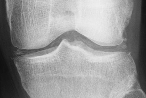
Plain radiography showed degenerative changes affecting the medial compartment of the knee and calcification of the menisci (Fig. 1a). The lateral view showed calcification within the origin of the medial head of gastrocnemius (Fig. 1b). CT revealed inhomogeneous deposition of calcium in the tendon (Fig. 2) and MRI confirmed the presence of a low-intensity lesion (Fig. 3). The symptoms were not relieved by the injection of 1% lidocaine into the knee.
A diagnosis of calcifying tendonitis was made. The symptoms did not improve after conservative management and decompression was performed four weeks later. A considerable amount of calcium deposit was removed at operation. Histological examination confirmed the diagnosis. There was dramatic relief from pain and she returned to work two months later.
Case 2. A 74-year-old female housekeeper presented in July 1998 with a history of post-traumatic posterior pain in the right knee for one week. There was marked tenderness in the region of the medial head of gastrocnemius. The range of flexion of the knee was from 30 deg to 90 deg.
Plain radiography showed calcification and CT confirmed that this was in the origin of the tendon of the medial head of gastrocnemius. A diagnosis of acute calcifying tendonitis was made and she obtained dramatic symptomatic relief after a local injection of 1% lidocaine and steroid.
Case 3. A 67-year-old female expert in the tea ceremony presented in October 1997 with a history of pain in the right knee for six months. There was marked tenderness in the region of the medial head of gastrocnemius and the range of flexion of the knee was from 20 deg to 95 deg. Plain radiography showed soft-tissue calcification which CT indicated to be within the origin of the tendon of the medial gastrocnemius. Over a period of eight months she was treated with local injections of steroid. The pain improved gradually but some limitation of movement persisted.
Discussion
Calcification in tendons is relatively common in CPPD crystal deposition, especially in tendo Achillis and the tendons of gastrocnemius and quadriceps.2 Yang et al3 reported an incidence of calcification in gastrocnemius of 31.9% (44/138) in patients with CPPD crystal deposition in the knee. The mean age of the patients was 72.6 years. These studies have dealt only with the radiological appearances of calcification with no reports of associated tendonitis.
Two of our patients had a probable diagnosis of CPPD crystal deposition according to the criteria of Ryan and McCarty.4
The cause of calcification within tendons is unknown. Suggested causes include hereditary, metabolic, post-traumatic and post-surgical conditions.5 Tendonitis induced by calcium deposition is well known in the rotator cuff5 and tendo Achillis.6
An inflammatory reaction within the tendon may be induced by the shedding of crystals from the deposits.7,8
In patients with tendonitis of gastrocnemius, there is marked tenderness in the region of the origin of the tendon of the medial head with posterior knee pain induced by stretching the tendon. An injection of 1% lidocaine with steroid into the tendon resulted in relief from pain and improvement in the range of movement of the knee.
Calcifying tendonitis is a differential diagnosis when calcification is found in relation to an osteoarthritic knee.
No benefits in any form have been received or will be received from a commercial party related directly or indirectly to the subject of this article.
References
1. Martel W, McCarter DK, Solsky MA, et al. Further observation of the arthropathy of calcium pyrophosphate dihydrate crystal deposition disease. Radiology 1981;141:1-15.
2. Pereira ER, Brown RR, Resnick D. Prevalence and patterns of tendon calcification in patients with chondrocalcinosis of the knee: radiologic study of 156 patients. Clin Imaging 1998;22:371-5.
3. Yang BY, Sartoris DJ, Resnick D, Clopton P. Calcium pyrophosphate dihydrate crystal deposition disease: frequency of tendon calcification about the knee. J Rheumatol 1996;23:883-8.
4. Ryan LM, McCarty DJ. Calcium pyrophosphate crystal deposition disease: pseudogout: articular chondrocalcinosis. In: McCarty DJ, ed. Arthritis and allied conditions: a textbook of rheumatology. Philadelphia: Lea & Febiger, 1989:1711-36.
5. Painter CF. Subdeltoid bursitis. Boston Med Surg J 1907;156:345-9.
6. Gerster JC, Lagier R, Boivin G. Achilles tendinitis associated with chondrocalcinosis. J Rheumatol 1980;7:82-8.
7. McCarty DJ Jr. Calcium pyrophosphate crystal deposition disease: pseudogout: articular chondrocalcinosis. In: McCarty DJ. ed. Arthritis and allied conditions: a textbook of rheumatology. Philadelphia: Lea & Febiger, 1972:040-60.
8. McCarty DJ. Calcium pyrophosphate dihydrate crystal deposition disease. Arthritis Rheum 1976;19:Suppl 3,275-85.
Y. Iguchi, N. Ihara, A. Hijioka, S. Uchida, T. Nakamura, A. Kikuta, T. Nakashima
From Akaike Kyodo Clinic and Chikuho Hospital, Fukuoka and the University of Occupational and Environmental Health, Kitakyushu, Japan
Y Iguchi, MD, Orthopaedic Surgeon
Department of Orthopaedic Surgery, Akaike Kyodo Clinic, 521-18 Akaike, Tagawa-gun, Fukuoka 822-1100, Japan.
N. Ihara, MD, Orthopaedic Surgeon
Department of Orthopaedic Surgery, Chikuho Hospital, 765-1 Yamabe, Nogata, Fukuoka 822-0034, Japan.
A. Hijioka, MD, Associate Professor
S. Uchida, MD, Assistant Professor
T. Nakamura, MD, Professor
Department of Orthopaedic Surgery
A. Kikuta, PhD, Professor
T. Nakashima, PhD, Assistant Professor
Department of Anatomy
University of Occupational and Environmental Health, 1-1 Iseigaoka, Yahatanishi-ku, Kitakyushu 807-8555, Japan.
Correspondence should be sent to Dr Y. Iguchi.
Copyright British Editorial Society of Bone & Joint Surgery Apr 2002
Provided by ProQuest Information and Learning Company. All rights Reserved


