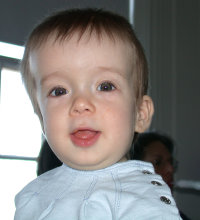
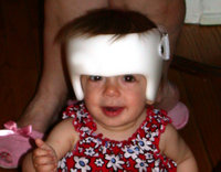
The birth of a child is an extremely exciting yet anxious time. Parents usually inspect their newborn infant as soon as possible after delivery and are happy after they determine that everything appears normal (eg, the infant has 10 fingers and 10 toes). Unfortunately, first impressions can lead to incorrect conclusions. Anatomical abnormalities and disease processes may present later in the newborn's life. For example, an abnormal head shape may not be evident at birth but can develop as the infant grows.
NORMAL CRANIAL ANATOMY
At birth, an infant's cranium is made up of eight bones:
* one ethmoidal,
* one frontal,
* one occipital,
* two parietal,
* one sphenoidal, and
* two temporal.
These cranial bones are separated by membranous spaces called sutures. The five cranial sutures are the frontal, coronal, lambdoidal, sagittal, and squamosal (Figure 1). The frontal suture usually closes fully by two to six years of age. If the frontal suture persists into adulthood, it is referred to as the metopic suture. The coronal, lambdoidal, sagittal, and squamosal sutures close clinically between six to 12 months of age but do not ossify completely until after 30 years of age. (1) All five sutures are flexible at birth and during early childhood for two reasons. First, the sutures allow the cranial bones to compress and override one another during the birth process. Distortion of the head often persists several days after birth; however, in most cases, this deformity resolves spontaneously during the rapid postnatal growth period. (2) Second, the sutures remain flexible during the first year of life to allow for normal skull expansion, which accommodates for brain growth. The neurocranium has achieved 63% of its growth at birth, 88% by one year of age, and 95% by 10 years of age. (3)
[FIGURE 1 OMITTED]
CRANIOSYNOSTOSIS
The premature fusion of one or more of the cranial sutures between the cranial bones is referred to as craniosynostosis. Premature fusion occurs in approximately one out of every 1,000 infants. Premature fusion may be associated with cranial and often facial skeletal anomalies. (4)
Craniosynostosis can be simple (ie, the fusion of one suture between two cranial bones) or complex (ie, the fusion of more than one suture). Cranial growth takes place at right angles to the direction of the suture; therefore, premature fusion of a suture results in growth arrest perpendicular to that suture. Compensatory growth, however, may occur in the direction of the suture. (5) characteristic head shape results depending on which suture or sutures are fused. This abnormal head shape is the first sign and the diagnostic determinant of craniosynostosis.
Most children are referred by their pediatrician to a pediatric neurosurgeon for possible neurosurgical intervention when a deformity progresses or fails to improve spontaneously. After an infant has been referred to a pediatric neurosurgeon, proper diagnosis is the critical first step in identifying appropriate interventions. Some patients with craniosynostosis also may have congenital syndromes. These syndromes must be identified so that treatment modalities can be chosen specifically for each individual patient.
Isolated sagittal synostosis, premature fusion of the sagittal suture, is the most common type of craniosynostosis and accounts for 56% to 58% of reported cases. (6) This condition usually occurs in full-term infants and typically is not associated with cognitive or psychomotor deficiencies. (7) The sagittal suture extends midline along the vertex of the cranium from the anterior coronal suture line to the posterior lambdoidal suture line. Due to its location, premature fusion of the sagittal suture is characterized by a long, narrow skull accompanied by characteristic frontal bossing, biparietal pinching, occipital protuberance, and a ridge at the sagittal suture (Figure 2). (8) The term scaphocephaly (ie, skiff- or keel-shaped head) is used to describe this common form of craniosynostosis. (9) This head shape results from the inability of the bone to grow perpendicular to the sagittal suture and from compensatory bone growth along the coronal and lambdoidal suture lines.
[FIGURE 2 OMITTED]
A diagnosis of sagittal synostosis is made after the characteristic head shape is noted in an otherwise healthy infant, and a thorough history and physical examination is obtained. As the infant continues to grow rapidly in the first few months of life, this unusual head shape becomes more apparent and easily recognized by the parents or the pediatrician, thereby necessitating a referral to a pediatric neurosurgeon. Skull x-rays, computerized tomography scans, and magnetic resonance imaging studies are not ordered routinely, as they rarely demonstrate any abnormality other than the sagittal suture fusion already noted by the neurosurgeon. (10) Sagittal synostosis does not resolve spontaneously or with less invasive, nonsurgical methods of correction (eg, molding helmets) because the sagittal suture is fused. The condition, therefore, requires surgical intervention. (11)
Pediatric neurosurgeons at Childrens Hospital Los Angeles (CHLA) evaluate an average of 15 to 20 children with sagittal synostosis per year. Surgical intervention is required in approximately 95% of these patients. Although surgery is the only treatment for this condition, parental decision and milder cases of sagittal synostosis account for the 5% nonsurgical intervention rate. Surgical correction of sagittal synostosis is performed mainly for cosmetic and neuropsychological considerations because isolated sagittal synostosis usually does not cause cognitive deficits or elevated intracranial pressure. A noncorrected unusual head shape, however, can result in significant psychological ramifications for a child, much like the disfigurement of a cleft lip. (12) The birth of a malformed infant also can precipitate a major family crisis, which disrupts the usual parent-infant bonding. (13) Surgical risks include infection, a poor cosmetic outcome, or bleeding that results in the need for a blood transfusion. Although the neurosurgeon considers the potential surgical risks versus the parents' desire for cosmetic outcome, in our society, correction has become the treatment of choice.
Infancy is the ideal time to treat craniosynostosis. (14) The optimal period for surgical correction of sagittal synostosis is three to six months of age; however, satisfactory correction still can be achieved if the patient presents after six months of age. (15) Age-related surgical outcome depends specifically on the rate and quality of cranial bone growth at the time of surgery. Infant bone is easily malleable making surgical manipulation less difficult. In addition, rapid cranial bone growth during the first six months of life results in maximal regrowth of cranial bone into the bony defects created by the surgery. This provides superior cosmetic outcomes. (16) Patients older than six months of age may require titanium or absorbable implants or bone replacement products to correct bony defects.
CALVARIAL VAULT REMODELING
At CHLA, surgical team members perform a calvarial vault remodeling (CVR) procedure to achieve optimal cosmetic results when correcting sagittal synostosis. Calvarial vault remodeling entails making a bicoronal incision in the scalp and removing, recontouring, and replacing cranial bone to produce a normal head shape. The goal of CVR for sagittal synostosis patients is to produce immediate and lasting correction of the deformity without any visible or palpable bony defect. (17) This goal is achieved by reducing the occipital protuberance and widening the parietal bones bilaterally to produce a normal shaped head.
Strip craniectomy is an alternative method of correction performed by neurosurgeons at other facilities. This more conservative procedure involves removing a narrow central strip of sagittal bone without recontouring or replacing the bone. Unfortunately, craniectomies become progressively less effective after six months of age. Although CVR is more involved and complicated, it produces significantly better cosmetic outcomes without additional complications other than the possible need for a blood transfusion. In addition, CVR can be used throughout infancy, making CVR the surgical procedure of choice at CHLA. (18)
PREOPERATIVE PHASE
The CVR procedure is scheduled as an elective surgery after the neurosurgeon and patient's parents or legal guardian decide that it is the preferred treatment intervention. Parents and family members are given the option to donate blood to the CHLA blood bank and direct its use for their infant's surgery because intraoperative blood loss is significant. Patients undergoing CVR receive at least one unit of packed red blood cells (PRBCs) intraoperatively. At CHLA, blood donated for a specific scheduled surgery must be donated at least 48 hours before the surgery to allow time for testing and processing. The PRBCs must be used within 35 days, or they are considered expired and not available for transfusion. (19)
Preoperative nursing assessment The patient and his or her parents report to the preoperative area the day before surgery for required laboratory testing and preoperative assessment and teaching. A complete blood count is drawn for baseline measurement as well as a blood type and cross-match. No other blood tests are required unless bleeding disorders are suspected. A nurse practitioner performs the preoperative history and physical examination. A perioperative nurse assesses the patient. Table 1 outlines the appropriate patient-specific nursing diagnoses and plan of care. Nursing diagnoses appropriate for this patient population include
* risk for ineffective family coping related to stress of surgery,
* risk for deficient fluid volume related to fluid volume loss, and
* risk for hypothermia related to exposure to cool environment, patient age, and exposed body surface.
Barring any abnormal findings, parents are given preoperative instructions, including requested hospital check-in time and NPO instructions, which depend on the age of the patient.
Preoperative teaching. The day of surgery, the circulating nurse and anesthesia care provider meet with the patient and his or her parents to provide more specific preoperative teaching. Informed consent is verified at this time. The circulating nurse confirms that the parents understand the donor directed blood transfusion process and verifies that the donor directed blood has been processed and is available for transfusion. The anesthesia care provider explains general anesthesia to the parents, including that
* the patient is induced using an inhalation induction in the OR;
* two IV lines are inserted after the patient is asleep to infuse fluid, medication, and blood; and
* an arterial line is inserted to monitor the patient's arterial blood pressure and provide access for drawing blood to test hematocrit levels intraoperatively when needed.
The nurse explains that the patient will be in the OR for approximately two to three hours. At CHLA, surgical time averages 126 minutes with additional time needed for induction, positioning, clipping the patient's hair if present, prepping, dressing application, and extubation. (20) The nurse reassures the parents that after surgery, the neurosurgeon will speak to them about how the procedure went and their child's condition.
The nurse explains that the child will be transferred to the postanesthesia care unit (PACU) after surgery when he or she is awake. The parents are informed that they will be allowed to be with their child after he or she has been settled and stabilized in the PACU. The nurse explains to the parents that their child's head shape will look fairly normal immediately after surgery; however, in a few hours, significant postoperative swelling will occur. Maximum swelling occurs in two or three days. Typically the swelling of the eyelids is such that the patient is unable to open his or her eyes temporarily. The parents are assured that the swelling, although uncomfortable, will not be terribly painful and will resolve with time. Pain medication will be given to the patient after surgery as needed. The nurse and anesthesia care provider ensure that all the parents' questions are answered before transferring the patient to the OR.
Operating room preparation. The circulating nurse is responsible for ensuring that the OR is prepared correctly. Before the patient is brought to the OR, the circulating nurse adjusts the temperature in the room to approximately 78 [degrees] F (25.6 [degrees] C) to accommodate for the patient's age and potential for intraoperative heat loss. The anesthesia care provider and circulating nurse ensure that warming lights are placed at an appropriate distance to help maintain optimal body temperature. They also provide a temperature regulating blanket that has been checked for proper functioning. This blanket can be applied to the patient after positioning to further prevent heat loss or to increase body temperature as needed. The circulating nurse places a sheepskin on the bed underneath the patient's body for padding as well as conservation of body heat. The circulating nurse prepares appropriately sized chest, hip, and ankle rolls for maintaining proper body alignment and to allow for adequate chest expansion while the patient is in the prone position. Finally, the circulating nurse checks the pediatric neurosurgical stabilizing device to ensure that all parts are available for head positioning.
INTRAOPERATIVE PHASE
The circulating nurse and anesthesia care provider transfer the patient to the OR. Premedication for separation anxiety usually is unnecessary for infants. The circulating nurse and anesthesia care provider place the patient in the supine position on the OR bed, and the anesthesia care provider induces general anesthesia using inhalation agents. The patient is intubated after the IV line has been placed. The arterial line then is inserted.
Positioning. The circulating nurse, neurosurgeon, and anesthesia care provider work cooperatively to place the patient in the prone position using a padded pediatric neurosurgical stabilizing device without fixating pins. Chest, hip, and ankle rolls with extra padding under the elbows and knees are used to reduce areas of pressure (Figure 3). An electrosurgical dispersive pad is applied properly, the patient is secured into place using safety straps, and the air-warming blanket is placed over the patient's body. The circulating nurse performs a final check to ensure proper body alignment and padding before beginning the prep.
[FIGURE 3 OMITTED]
Prep. The neurosurgeon clips the patient's hair, if present, in a wide bicoronal strip surrounding the area where the incision will be made. The bicoronal incision line then is identified using a surgical marking pen. The anesthesia care provider administers prophylactic antibiotics, if ordered by the neurosurgeon. Cefazolin 25 mg per kg is the antibiotic of choice, with substitutions made if allergies exist. The circulating nurse preps the head in a circular manner proceeding from the incision line circumferentially outward, after which the surgeon and scrub person position the surgical drapes.
Surgical procedure. The neurosurgeon makes a bicoronal scalp incision extending from ear to ear using a bilateral zigzag incision pattern in the temporal areas. This incision pattern minimizes the visibility of the scar postoperatively because it allows hair regrowth to more effectively cover the incision. The patient's hair will fall down and forward with gravity, crossing the scar rather than exposing the area of alopecia along a vertical scar.
After the incision is made, the anesthesia care provider begins a blood transfusion, which continues slowly throughout the procedure. The anesthesia care provider tests arterial blood gases periodically and keeps the neurosurgeon informed throughout the procedure regarding the patient's response to blood loss.
Using the same bicoronal incision, the neurosurgeon retracts the scalp to expose the cranium from the coronal suture to the lambdoidal suture. The dura mater is kept intact throughout the entire procedure. The neurosurgeon uses a pneumatic craniotome to remove a 2-cm to 3-cm central strip of bone containing the sagittal suture (Figure 4). The strip extends from the coronal suture to the lower occipital region. The scrub person places the bone strip on the back table to be recontoured and collects any bone dust from the use of the pneumatic craniotome for later replacement.
[FIGURE 4 OMITTED]
The neurosurgeon makes perpendicular, barrel-staving cuts in the parietal bones along the length of the central sagittal strip extending temporally to the squamosal sutures (Figure 5). These barrel-staving cuts allow for widening and curving of the parietal bones to create a more normal head shape. The neurosurgeon recontours the piece of removed central sagittal strip using a bone-contouring rongeur. Bone fragments are set aside for later replacement.
[FIGURE 5 OMITTED]
The neurosurgeon places the recontoured strip into the now widened midline bony detect and secures it in place typically using 4-0 absorbable sutures. The neurosurgeon reduces the occipital protuberance by securing the central sagittal strip to the occipital bones using nonabsorbable sutures or 28-g surgical steel wire. The neurosurgeon drills small holes into both bones to provide access for the wire to secure the two bones together. This results in a more rounded head shape.
After the central sagittal strip has been replaced and secured, small bony defects or gaps are present where cuts in the bone have been made. The neurosurgeon uses the remaining bone fragments from the cutting and recontouring of the central strip and the bone dust collected from the use of the pneumatic craniotome to partially fill in the spaces between bone cuts. At the time of closure, small bony defects will exist. These defects will fill in postoperatively with cranial bone regrowth because of the rapid bone growth associated with infants.
The scalp then is reapproximated and closed using two layers of absorbable suture after all the bone is secure. Surgical drains are needed infrequently. The wound is dressed with gauze, which is taped into place. A headwrap dressing is avoided so the patient is less likely to be uncomfortable when scalp swelling develops. After the dressing is placed, surgical team members carefully reposition the patient supine and the anesthesia care provider extubates the patient.
POSTOPERATIVE PHASE
Immediately after extubation, the patient is transferred to a stretcher or crib and transported to the PACU. The PACU nurse assigned to the patient focuses primarily on postanesthesia care, which includes monitoring and documenting
* airway management,
* temperature regulation,
* pain control,
* vital signs,
* oxygen saturation levels, and
* continuous electrocardiogram (ECG) readings.
If the patient is stable, arterial blood pressure readings via the arterial line are not monitored. The PACU nurse draws a hematocrit level. If the hematocrit level is acceptable to the neurosurgeon (ie, ideally above 40%), the arterial line is discontinued before the patient is transferred to the inpatient surgical unit. Intravenous morphine sulfate 0.05 mg per kg to 0.1 mg per kg is the medication of choice for immediate and short-term postoperative pain control. Typically, patients receive morphine sulfate every two to three hours as necessary for pain control for 48 hours postoperatively.
The patient's parents are allowed to visit in the PACU after approximately 30 minutes if the patient is stable and recovering well from anesthesia. This decreases both patient and parent anxiety. Postanesthesia care unit staff members transfer the patient by stretcher or crib to the inpatient surgical unit after initial recovery in the PACU (ie, at least one hour) if the patient remains stable. On the inpatient unit, routine nursing care is performed following CHLA post-operative protocols and physicians' orders. The patient is administered prophylactic antibiotics for 24 hours and has hematocrit levels monitored every day until discharge. Acetaminophen with codeine elixir is administered for pain control after the initial 48 hours for mild discomfort and is prescribed for home use after discharge. The average hospital stay at CHLA for patients having undergone CVR is 4.4 days. (21)
The patient and his or her parents are encouraged to return to normal activities after discharge from the hospital. Parents are instructed to avoid getting the head dressing wet. Patients have a follow-up visit with the neurosurgeon at one week and eight weeks postoperatively. These postoperative office visits allow the neurosurgeon to assess the skull bones during the healing process and evaluate the skull defects and regrowth.
COMPLICATIONS
Fortunately, complications after CVR are infrequent. In a 10-year period, no permanent complications were encountered by patients who underwent CVR at CHLA. (22) The two complications most likely to occur are postoperative infection and the need for a blood transfusion.
Postoperative infection& Postoperative infections are rare. Prophylactic IV antibiotics are administered preoperatively before the initial incision is made. Twenty-four hours of postoperative IV antibiotics also are ordered. This antibiotic protocol greatly reduces the incidence of postoperative infection.
Blood transfusions. Precautions are taken to maintain optimal hemodynamics perioperatively so the need for a blood transfusion is considered a surgical complication. Blood transfusions are necessary in CVR surgery, however, due to the constant loss of blood during the procedure, especially considering the smaller blood volume of an infant (ie, 75 to 80 mL per kg). A hematocrit level of 45% is the goal at the close of the patient's incision. To achieve this, an average of 357 mL of PRBCs are transfused during a typical surgical procedure. (23) Typically in the first 24 postoperative hours, the hematocrit level drops 10% with a further drop after 24 hours. Additional postoperative blood transfusions are unlikely if the patient's hematocrit level is 45% at the end of the surgical procedure.
CASE STUDY
Billy is a four-month-old otherwise healthy caucasian male who presented with sagittal synostosis for a CVR procedure. Mr and Mrs K, Billy's parents, were present with him before surgery in the preoperative holding area. The circulating nurse had verified the availability of one unit of donor-directed PRBCs for use during the procedure. The circulating nurse, along with the anesthesia care provider, visited with the patient and his parents preoperatively. Billy appeared healthy without any signs of acute illness. After discussing CVR and perioperative expectations with Mr and Mrs K, the circulating nurse and anesthesia care provider transferred Billy to the OR.
In the OR, a pulse oximeter, ECG leads, and a blood pressure cuff were applied to Billy. The anesthesia care provider induced Billy under general anesthesia via mask and appropriate anesthetic gases. Further anesthetic agents were administered via an IV line that was inserted after Billy was anesthetized. An appropriately sized endotracheal tube was inserted and secured in place. A second IV line and an arterial line were inserted as well.
The neurosurgeon, anesthesia care provider, and circulating nurse worked cooperatively using the log roll technique to turn Billy from the supine to the prone position. Billy was placed on sheepskin and positioned with rolls so that his head rested on the neurosurgical stabilizing device. All team members focused on maintaining Billy's proper body alignment and protecting his endotracheal tube and vascular access during positioning. The circulating nurse then placed the electrosurgical dispersive pad, safety strap, and temperature regulating blanket on Billy.
The neurosurgeon shaved Billy's head, and the nurse bagged and labeled the hair. Billy's clipped hair was saved and given to his parents because it is considered patient property. The anesthesia care provider administered prophylactic antibiotics as the neurosurgeon prepped the patient. The surgeon made a zigzag incision.
The anesthesia care provider tested Billy's arterial blood gas. Billy's baseline hematocrit level was 30%, which is normal for a four-month old child. The anesthesia care provider began a slow transfusion of the donor-directed PRBCs. Throughout the procedure, two other blood gases were tested and a 44% hematocrit level was measured during wound closure. Billy received the entire unit of donor-directed PRBCs equaling 340 mL. The wound was dressed using a petroleum gauze strip, 4 x 4 gauze, and a manufactured dressing sheet. Billy was repositioned carefully into the supine position and extubated.
After a safe extubation, Billy immediately was transferred via stretcher to the PACU. Billy's vital signs were monitored, and they remained stable throughout the entire PACU stay. After Billy was in the PACU for 30 minutes, Mrs K was allowed to enter and stay at Billy's side. Billy required one dose of 0.05 mg per kg of morphine sulfate via IV in the PACU. Fifteen minutes before Billy's transfer to the inpatient surgical unit, the arterial line was discontinued. When he was comfortable, the PACU nurse transferred Billy to the inpatient pediatric surgical unit.
After 36 hours, pain control was maintained using acetaminophen with codeine. Billy received 24 hours of antibiotics and required no further blood transfusions. Billy's hematocrit level stabilized at 31% by postoperative day four. He was discharged home eating well, and his parents were comfortable with his postoperative home care needs. Billy was seen by the neurosurgeon at one week and eight weeks postoperatively for routine office visits. The bony defects in his skull filled in without any abnormalities. Billy's postoperative phase proved uneventful, and his parents were pleased with the surgical results.
CONCLUSION
Parents are surveyed postoperatively to evaluate their satisfaction with the cosmetic results of the CVR procedure performed at CHLA. The survey results are very positive with 79% of parents considering their child's cosmetic outcomes as excellent. Excellent is defined by the parents as resulting in good to excellent correction with no visible or palpable abnormality. (24)
The author would like to thank Sooho Choi, MD, for his assistance with this article.
NOTES
(1.) S A Wolfe, S Berkowitz, Plastic Surgery of the Facial Skeleton (Boston: Little, Brown and Co, 1989) 489.
(2.) Ibid.
(3.) Ibid.
(4.) D Marchac, D Renier, "Treatment of craniosynostosis in infancy," Clinics in Plastic Surgery 14 (January 1987) 61-72.
(5.) Wolfe, Berkowitz, Plastic Surgery of the Facial Skeleton, 489.
(6.) J M Pensler, S J Ciletti, T Tomita, "Late correction of sagittal synostosis in children," Plastic and Reconstructive Surgery 97 (June 1996) 1362-1370.
(7.) M H Huang et al, "The differential diagnosis of abnormal head shapes: Separating craniosynostosis from positional deformities and normal variants," The Cleft Palate--Craniofacial Journal 35 (May 1998) 204-211.
(8.) Pensler, Ciletti, Tomita, "Late correction of sagittal synostosis in children," 1362-1370.
(9.) Ibid.
(10.) T A Maugans, J G McComb, M Levy, "Surgical management of sagittal synostosis: A comparative analysis of strip craniectomy and calvarial vault remodeling," Pediatric Neurosurgery 27 (September 1997) 137-148.
(11.) Huang et al, "The differential diagnosis of abnormal head shapes: Separating craniosynostosis from positional deformities and normal variants," 204-211.
(12.) F A Boop, K Shewmake, W M Chadduck, "Synostectomy versus complex cranioplasty for the treatment of sagittal synostosis," Child's Nervous System 12 (July 1996) 371-375.
(13.) M M Cohen, Craniosynostosis: Diagnosis, Evaluation, and Management (New York: Raven Press, 1986) 32-34.
(14.) Marchac, Renier, "Treatment of craniosynostosis in infancy," 61-72.
(15.) Maugans, McComb, Levy, "Surgical management of sagittal synostosis: A comparative analysis of strip craniectomy and calvarial vault remodeling," 137-148.
(16.) Marchac, Renier, "Treatment of craniosynostosis in infancy," 61-72.
(17.) Maugans, McComb, Levy, "Surgical management of sagittal synostosis: A comparative analysis of strip craniectomy and calvarial vault remodeling," 137-148.
(18.) Ibid.
(19.) M Brown, P K Whalen, "Red blood cell transfusion in critically ill patients," Critical Care Nurse, http://www.aacn.org/aacn/jrnlccn.nsf (accessed 26 June 2001).
(20.) Maugans, McComb, Levy, "Surgical management of sagittal synostosis: A comparative analysis of strip craniectomy and calvarial vault remodeling," 137-148.
(21.) Ibid.
(22.) Ibid.
(23.) Ibid.
(24.) Ibid.
Susan A. Johnston, RN, BSN, CNOR, is the specialty leader for neurosurgery at Childrens' Hospital Los Angeles.
COPYRIGHT 2001 Association of Operating Room Nurses, Inc.
COPYRIGHT 2002 Gale Group


