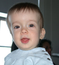
An image of a child with Lamdoid Cranial Synostosis. Notice the swelling on the right side of the head.A child wearing a cranial band.
Find information on thousands of medical conditions and prescription drugs.

Craniosynostosis

Craniosynostosis is a medical condition in which the sutures in the skull of an infant close too early, causing problems with normal brain and skull growth. It can result in craniostenosis, which is the skull deformity caused by the premature closure of the cranial sutures. Also intracranial pressure can be increased. more...

Home
Diseases
A
B
C
Angioedema
C syndrome
Cacophobia
Café au lait spot
Calcinosis cutis
Calculi
Campylobacter
Canavan leukodystrophy
Cancer
Candidiasis
Canga's bead symptom
Canine distemper
Carcinoid syndrome
Carcinoma, squamous cell
Carcinophobia
Cardiac arrest
Cardiofaciocutaneous...
Cardiomyopathy
Cardiophobia
Cardiospasm
Carnitine transporter...
Carnitine-acylcarnitine...
Caroli disease
Carotenemia
Carpal tunnel syndrome
Carpenter syndrome
Cartilage-hair hypoplasia
Castleman's disease
Cat-scratch disease
CATCH 22 syndrome
Causalgia
Cayler syndrome
CCHS
CDG syndrome
CDG syndrome type 1A
Celiac sprue
Cenani Lenz syndactylism
Ceramidase deficiency
Cerebellar ataxia
Cerebellar hypoplasia
Cerebral amyloid angiopathy
Cerebral aneurysm
Cerebral cavernous...
Cerebral gigantism
Cerebral palsy
Cerebral thrombosis
Ceroid lipofuscinois,...
Cervical cancer
Chagas disease
Chalazion
Chancroid
Charcot disease
Charcot-Marie-Tooth disease
CHARGE Association
Chediak-Higashi syndrome
Chemodectoma
Cherubism
Chickenpox
Chikungunya
Childhood disintegrative...
Chionophobia
Chlamydia
Chlamydia trachomatis
Cholangiocarcinoma
Cholecystitis
Cholelithiasis
Cholera
Cholestasis
Cholesterol pneumonia
Chondrocalcinosis
Chondrodystrophy
Chondromalacia
Chondrosarcoma
Chorea (disease)
Chorea acanthocytosis
Choriocarcinoma
Chorioretinitis
Choroid plexus cyst
Christmas disease
Chromhidrosis
Chromophobia
Chromosome 15q, partial...
Chromosome 15q, trisomy
Chromosome 22,...
Chronic fatigue immune...
Chronic fatigue syndrome
Chronic granulomatous...
Chronic lymphocytic leukemia
Chronic myelogenous leukemia
Chronic obstructive...
Chronic renal failure
Churg-Strauss syndrome
Ciguatera fish poisoning
Cinchonism
Citrullinemia
Cleft lip
Cleft palate
Climacophobia
Clinophobia
Cloacal exstrophy
Clubfoot
Cluster headache
Coccidioidomycosis
Cockayne's syndrome
Coffin-Lowry syndrome
Colitis
Color blindness
Colorado tick fever
Combined hyperlipidemia,...
Common cold
Common variable...
Compartment syndrome
Conductive hearing loss
Condyloma
Condyloma acuminatum
Cone dystrophy
Congenital adrenal...
Congenital afibrinogenemia
Congenital diaphragmatic...
Congenital erythropoietic...
Congenital facial diplegia
Congenital hypothyroidism
Congenital ichthyosis
Congenital syphilis
Congenital toxoplasmosis
Congestive heart disease
Conjunctivitis
Conn's syndrome
Constitutional growth delay
Conversion disorder
Coprophobia
Coproporhyria
Cor pulmonale
Cor triatriatum
Cornelia de Lange syndrome
Coronary heart disease
Cortical dysplasia
Corticobasal degeneration
Costello syndrome
Costochondritis
Cowpox
Craniodiaphyseal dysplasia
Craniofacial dysostosis
Craniostenosis
Craniosynostosis
CREST syndrome
Cretinism
Creutzfeldt-Jakob disease
Cri du chat
Cri du chat
Crohn's disease
Croup
Crouzon syndrome
Crouzonodermoskeletal...
Crow-Fukase syndrome
Cryoglobulinemia
Cryophobia
Cryptococcosis
Crystallophobia
Cushing's syndrome
Cutaneous larva migrans
Cutis verticis gyrata
Cyclic neutropenia
Cyclic vomiting syndrome
Cystic fibrosis
Cystinosis
Cystinuria
Cytomegalovirus
Dilated cardiomyopathy
Hypertrophic cardiomyopathy
Restrictive cardiomyopathy
D
E
F
G
H
I
J
K
L
M
N
O
P
Q
R
S
T
U
V
W
X
Y
Z
Medicines

Normal skull development

In humans, the adult skull is normally made up of 28 bones. Except for the mandible, all of the bones of the skull are joined together by sutures, rigid articulations permitting very little movement.

At birth, the human skull is made up of 45 separate bony elements. As growth occurs, many of these bony elements gradually fuse together into solid bone (for example, the frontal bones).

The bones of the roof of the skull are initially separated by regions of dense connective tissue. At birth these regions are fibrous and moveable, necessary for birth and later growth. Larger regions of connective tissue, called fontanelles, occur where certain bony elements meet. As growth and ossification progress, the connective tissue of the fontanelles is invaded and replaced by bone. The posterior fontanelle usually closes by eight weeks, but the anterior fontanelle can remain up to eighteen months.

Pathophysiology

When one or more sutures fuse prematurely, skull growth can be restricted perpendicular to the suture. If multiple sutures fuse while the brain is still increasing in size, intracranial pressure can increase.

Primary craniosynostosis is believed to be a result of primary defect in the mesenchymal layer ossification in the cranial bones. Secondary craniosynostosis is a result of primary failure of brain growth.

Treatment

Surgery is typically used to seperate the fused sutures of the skull as well as to reshape the skull. Without surgery, blindness and mental retardation is a typical outcome. To treat the cosmetic troubles, a combination of orthodontic and orthognathic surgery can be used to relieve some of the midface deficiency.

Typical surgery begins with a zigzag incision from ear to ear across the top of the head. The scar left by this type of incision makes the hair look more natural than that left by a straight incision would. Raney clips are typically used to curtail bleeding, as cauterization would not result in an aesthetically pleasing result upon healing. Once the scalp is peeled back, pilot holes are drilled through the skull. These pilot holes are then connected, separating the skull into several pieces. Once reshaped, these pieces are placed back on the head (typically in an altered configuration) and held together by a combination of dissolving sutures, plates, and screws. These plates and screws are typically composed of a copolymer comprised of polyglycolic and polylactic acid and will break down into water and carbon dioxide within a year. Demineralized bone matrix is often used to fill gaps left by the expanded skull. Once the Raney clips are removed, dissolving sutures are again used to close the incision.

Read more at Wikipedia.org


[List your site here Free!]


Cigarette smoking - Drugs, Pregnancy, And Lactation
From OB/GYN News, 10/1/03 by Gerald G. Briggs

About 15%-29% of pregnant women smoke in pregnancy, which can cause significant fetal and infant toxicity and lead to other adverse outcomes that may persist long after birth.

Many physicians aren't aware of all the problems associated with smoking before or during gestation, including reduced fecundity, ectopic (tubal) pregnancy, spontaneous abortion, abnormal placentation, growth retardation, premature rupture of the membranes (PROM), preterm delivery, and increased perinatal mortality

The association of smoking with spontaneous abortion has been controversial. The overall risk appears to be only slightly increased, but may be increased twofold in heavy smokers.

The risk of placenta previa and placental abruption is increased by about 50%, rising with the number of cigarettes smoked.

Symmetrical fetal growth retardation is a well-known adverse effect of maternal smoking during pregnancy, with an average reduction in birth weight of 200 g. The risk of having a low-birth-weight (<2,500 g) infant is doubled among smokers, and birth length and head circumference are reduced.

Stopping smoking before 32 weeks' gestation may prevent the adverse effects on birth weight and head circumference but has not been shown to prevent decreased height or increased ponderal index (i.e., a shorter and/or fatter infant).

For women who smoke at least 20 cigarettes/day, there is a twofold increased risk of PROM before 33 weeks. About 5% of all premature births can be attributed to maternal smoking.

If all smoking during pregnancy were eliminated, perinatal mortality, would be reduced by an estimated 10%. The increased risk of perinatal mortality is at tributable to low birth weight, prematurity, and abnormal placentation.

Accumulating evidence indicating that smoking during pregnancy in creases the risk of sudden infant death syndrome (SIDS) includes two recent studies which found a 3.5-fold greater risk of SIDS among babies of smokers.

Whether smoking induces congenital malformations is controversial, because studies over the past 20 years have shown both positive and negative associations between smoking and birth defects. While such an association may not exist, some recent evidence suggests that the conflicting results may be due to underreporting of smoking in pregnancy, unrecognized confounding variables, or interactions with genetic or environmental factors.

In general, studies have indicated that the risk of cleft lip and palate associated with smoking--if it exists at all--is low, with an odds ratio <2. The lack of consistency among studies concerning these defects suggests there may be an interaction between smoking and genetic factors. Recent data have suggested that maternal smoking increases the risk of foot deformities by a factor of 1.3-2.4. Risk increases more than 20-fold when combined with family history. These results are thought to be consistent with an interaction between a genetic factor and smoking.

Associations between maternal smoking and isolated craniosynostosis have been reported.

Transverse limb reduction defects were associated with maternal smoking in other studies, with the strongest association--an odds ratio of 2--when both parents smoked. Moderately significant associations between maternal smoking and urinary tract abnormalities in infants have recently been reported. There was no dose-response effect, possibly because of an increase in spontaneous abortions and stillbirths that may have lowered the number of live-born infants with these defects.

Conotruncal heart defects--such as tetralogy of Fallot, ventricular septal defects, and transposition of the great arteries--have been associated with maternal smoking, with rates about twice those of nonsmokers. These associations are biologically plausible, because these defects are thought to be related to vascular disruption.

Maternal smoking appears to be related to neurodevelopmental abnormalities, such as idiopathic mental retardation in children with no evidence of CNS damage or other developmental disabilities. Maternal smoking has also been related to behavioral problems and auditory deficits in the offspring.

In utero exposure to smoking has been associated with impaired neonatal lung function. And retinal vessel abnormalities, arterial narrowing/straightening, and venous dilatation/tortuosity were markedly increased in the eyes of neonates of smoking mothers, compared with those of nonsmoking mothers, but resolved by 6 months.

GERALD G. BRIGGS, B. PHARM., is pharmacist clinical specialist, Women's Hospital, Long Beach Memorial Medical Center; clinical professor of pharmacy, University of California, San Francisco; and adjunct professor of pharmacy, University of Southern California, Los Angeles. He is also coauthor of the reference book "Drugs in Pregnancy and Lactation."

COPYRIGHT 2003 International Medical News Group
COPYRIGHT 2003 Gale Group

Return to Craniosynostosis
Home Contact Resources Exchange Links ebay