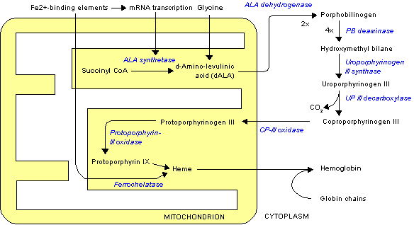
The patient was a 35-year-old, white woman with a history of rapidly progressive liver cirrhosis diagnosed 1 month before her most recent admission. On this admission, she had signs and symptoms consistent with acute liver failure, hepatic encephalopathy, and recurrent supraventricular tachycardia.
The patient reported a history of tachycardia of unknown origin since the age of 17 years for which she was taking digoxin (Lanoxin). One or 2 years ago bouts of tachycardia became more frequent and atenolol (Tenormin) was added to her drug regimen. Electrocardiogram showed right axis deviation with T-wave inversions in V^sub 1^ through V^sub 3^.
The patient had 3 siblings. Two of her sisters had histories of cardiac arrhythmias. Her younger brother died from a fatal cardiac arrhythmia during a surgical procedure. The patient's medical history was significant for recurrent abdominal pain, which first began at the age of 14 years and had continued since that time. She had been hospitalized on several occasions for the abdominal pain and undergone various surgical procedures to determine the cause of the pain, but to no avail. During the last 12 months, the pain had become severe and sharp in nature. A diagnosis of hereditary coproporphyria was eventually established. She received infusions of glucose and hematin, which gave her symptomatic relief. Her hospital stay was complicated by worsening liver, renal, and respiratory failure with bilateral pulmonary edema and pleural effusions. She died from a fatal cardiac arrhythmia 10 days after the admission.
At autopsy, cardiomegaly with right atrial and ventricular dilation was identified. The thickness of the fatty replacement ranged from 15 (at the basal region) to 8 mm (adjacent to the apex). The right ventricular myocardial wall exhibited a full-thickness replacement of myocardium by mature adipose tissue intermingled with focal bundles of the remaining myocardium (Figure 1). Histologic examination of the right and left ventricles confirmed gross findings. Myocytes persisted only as scattered isolated cells or small strands among the fat cells of the free right ventricular wall (Figure 2). The right ventricle had a thickness of 8 mm and the left ventricle had a thickness of 19 mm. The cut surface of the posterior wall of the left ventricular myocardium exhibited an area of total replacement of the myocardium by adipose tissue (Figure 3).
What is your diagnosis?
Pathologic Diagnosis: Fatty Variant of Arrhythmogenic Right and Left Ventricular Cardiomyopathy Associated With Hereditary Hepatic Coproporphyria
Fatty variant of arrhythmogenic right and left ventricular cardiomyopathy (ARVC) is a familial cardiomyopathy histologically characterized by adipose or fibroadipose replacement of myocytes.
Its prevalence is estimated to be 1 in 5000 people.1 In Italy, it is the most common cause of sudden death in young people. In the United States, it accounts for 17% of all sudden death in young people.2 Autosomal dominant inheritance has been reported in approximately 30% of cases.' In addition, ARVC has a wide genetic, clinical, and histologic spectrum. Although no gene has yet been discovered, several chromosomal loci have been mapped: 14q23, 1q42, 14q12, 2q32, 3p23, 10p12, and 17q21.
It is well recognized that ARVC is a disease with highly variable clinical manifestations, ranging from those that are asymptomatic and slowly progressive to more severe disease presenting with sudden unheralded death in young individuals. Some patients have a normal life expectancy, with the diagnosis being made only post mortem.
The main clinical manifestations of the disease are arrhythmias, complete heart block, heart failure, and sudden death.3 Arrhythmias that characterize ARVC include idiopathic ventricular fibrillation, ventricular extrasystoles, supraventricular tachycardia,4 and ventricular tachycardia of right ventricular origin (with a left bundle branch pattern). The typical electrocardiographic abnormality is T-- wave inversion in leads V^sub 1^ through V^sub 3^.3 The right ventricle is most frequently involved, but left-sided or biventricular cardiac disease also occurs.5 Two pathologic types of ARVC have been proposed: a variant of ARVC characterized exclusively by fat replacement and a variant with fibrofatty replacement of myocytes.
Although extensively discussed in the literature, the origin and natural history of this condition are still unclear.
The polymorphism of this disorder appears to be the result of several basic characteristics of the heart musculature: deposition of fat in the heart musculature, replacement of myocardium by fat, and susceptibility to environmental factors.
Several theories of the origin and pathogenesis of ARVC have been advanced.
According to the diontogenic theory, the absence of myocardium is thought to be the consequence of a congenital aplasia or hypoplasia of the right ventricular wall similar to the parchment heart characterized by gross cardiac structural defect present at birth, which was described by Uhl in 1952. The use of the term arrhythmogenic right ventricular dysplasia (maldevelopment) is in agreement with this theory. It is now recognized that Uhl anomaly and ARVC are 2 separate entities.
Myocellular transdifferentiation can be an alternative mechanism to fatty replacement of lost myocytes in the pathogenesis of ARVC. Gene defect mapped to chromosome 14q23-q246 favors genetically determined atrophy similar to the one that is well described in the skeletal muscle of patients with Duchenne and Becker diseases. The 14q-q24 region includes the genes of Beta-spectrin and alphaactinin. The similarity between the myocardial dystrophy observed in ARVC and the skeletal muscular dystrophy observed in Duchenne and Becker diseases and the structural homology between the alpha-actinin gene and the aminoterminal domain of dystrophin are all highly suggestive of a defective alpha-actinin gene. If this theory proves to be true, then the term arrhythmogenic right ventricular dystrophy might appear the most appropriate.
In the inflammatory theory, the fibrofatty replacement is regarded as a consequence of reparative process in the setting of chronic myocarditis. A genetic predisposition to viral infection eliciting immune reactions cannot be excluded. The finding of lymphocytic infiltrates has led to the consideration of ARVC as a chronic myocarditis. Various infectious agents, such as Tryponosoma cruzi, rubella virus, and mycoplasma, have been related to ARVC. Coxsackie virus genome in the myocardium of patients with ARVC was identified using reverse transcriptase-nested polymerase chain reaction protocol and nucleic acid sequencing.7
In the degenerative theory, the loss of myocytes is considered to be a result of progressive myocyte loss due to some metabolic or ultrastructural defect, resulting in substitution of degenerated myocytes by fat.
It was recently suggested that myocardial cell death in ARVC could represent a programmed cell death known as apoptosis.8
Clinical diagnosis can be established based on the presence of minor and major criteria encompassing structural, functional, histologic, cardiographic, and genetic factors according to the International Society and Federation of Cardiology.
References
1. Thiene G, Basso C, Danieli G, Rampazzo A, Corrado D, Nava A. Arrhythmogenic right ventricular cardiomyopathy. Trends Cardiovasc Med. 1997;7:8490.
2. Chen WK, Edwards WD, Hummill SC, Bailey KR, Ballard DJ, Gersh BJ. Sudden unexpected non-traumatic death in 54 young adults: a 30-year population-based study. Am J Cardiol. 1995;76:148-152.
3. Kullo Ii, Edwards WD, Seward 113. Right ventricular dysplasia: the Mayo Clinic experience. Mayo Clin Proc. 1995;70:541-548.
4. Tone IL, Castro-Miranda R, Iwa T, Poulain F, Frank R, Fontaine GH. Frequency of supraventricular tachyarrhythmias in arrhythmogenic right ventricular dysplasia. Am J Cardiol. 1991;67:1153.
5. Pinamonti B, Pagnan L, Bussani R, Ricci C, Silvestri F, Camerini F. Right ventricular dysplasia with biventricular involvement. Circulation. 1998;98:19431945.
6. Rampazzo A, Nava A, Danieli C, et al. The gene for arrhythmogenic right ventricular cardiomyopathy maps to chromosome 14q23-q24. Hum Mol Genet. 1994;3:2151-2154.
7. Grumbach IM, Vonhof S, Stille-Siegener M, et al. Coxsackievirus genome in myocardium of patients with arrhythmogenic right ventricular dysplasia/cardiomyopathy. Cardiology. 1998;89:241-245.
8. Mallat Z, Tedgui A, Fontaliran F, Frank R, Durigon M, Fontaine G. Evidence of apoptosis in arrhythmogenic right ventricular dysplasia. N EnglJ Med. 1996; 335:1190-1195.
Irina Mikolaenko, MD; Micheal G. Conner, MD
Accepted for publication September 10, 2001.
From the Department of Pathology, Division of Anatomic Pathology, University of Alabama at Birmingham, Birmingham, Ala.
Corresponding author: Michael G. Conner, MD, Department of Pathology, Division of Anatomic Pathology, University of Alabama at Birmingham, 506 Krake Bldg, 619 S 19th St, Birmingham, AL 35233-6823 (e-mail: mgconner@path.uab.edu).
Copyright College of American Pathologists Jun 2002
Provided by ProQuest Information and Learning Company. All rights Reserved


