Hereditary hemochromatosis
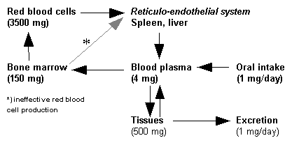
Haemochromatosis, also spelled hemochromatosis, is a hereditary disease characterized by improper processing by the body of dietary iron which causes iron to accumulate in a number of body tissues, eventually causing organ dysfunction. It is the main iron overload disorder. more...
Signs and symptoms
Haemochromatosis is notoriously protean, i.e. it presents with symptoms that are often initially attributed to other diseases.
Symptoms may include:
- Malaise
- Liver cirrhosis (with an increased risk of hepatocellular carcinoma, affecting up to a third of all homozygotes) - this is often preceded by a period of a painfully enlarged liver.
- Insulin resistance (often patients have already been diagnosed with diabetes mellitus type 2)
- Erectile dysfunction and hypogonadism
- Congestive heart failure, arrhythmias or pericarditis
- Arthritis of the hands (MCP and PIP joints), knee and shoulder joints
- Dysfunction of certain endocrine organs:
- Pancreatic gland
- Adrenal gland (leading to adrenal insufficiency)
- Parathyroid gland (leading to hypocalcaemia)
- Pituitary gland
- Testes or ovary (leading to hypogonadism)
- A darkish colour to the skin (see pigmentation, hence its name Diabete bronze when it was first described by Armand Trousseau in 1865)
- An increased susceptibility to certain infectious diseases caused by:
- Vibrio vulnificus infections from eating seafood
- Listeria monocytogenes
- Yersinia enterocolica
- Salmonella enteritidis (serotype Typhymurium)
- Klebsiella pneumoniae
- Escherichia coli
- Rhizopus arrhizus
- Mucor species
Males are usually diagnosed after their forties, and women about a decade later, owing to regular iron loss by menstruation (which ceases in menopause).
Diagnosis
Imaging features
Clinically the disease may be silent, but characteristic radiological features may point to the diagnosis. The increased iron stores in the organs involved, especially in the liver and pancreas, result in an increased attenuation at unenhanced CT and an decreased signal intensity at MR imaging. Haemochromatosis arthropathy includes degenerative osteoarthritis and chondrocalcinosis. The distribution of the arthropathy is distinctive, but not unique, frequently affecting the second and third metacarpophalangeal joints of the hand.
Chemistry
A first step is the measurement of ferritin, the tissue form of accumulated iron which is shed into the blood. Other markers of iron metabolism are the iron carrying protein transferrin (decreased), transferrin saturation (increased) and serum iron (increased). Genetic testing is performed if the biochemical markers are deranged.
Other blood tests routinely performed: blood count, renal function, liver enzymes, electrolytes, glucose (and/or an oral glucose tolerance test (OGTT)).
Read more at Wikipedia.org


