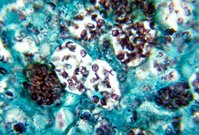
Disseminated histoplasmosis is a serious disease that affects the skin, lungs, and internal organs. It is one of the diseases that characterize acquired immunodeficiency syndrome (AIDS), and in endemic areas is one of the more commonly observed infections in AIDS patients. The mortality rate in patients with AIDS and histoplasmosis is high if untreated. Disseminated histoplasmosis may have a variety of dermatological manifestations. In this article, we provide the first report of diffuse ulcerations due to disseminated histoplasmosis. These ulcers developed while the patient was on stavudine, lamivudine, and indinavir, and had a CD-4 count of 525 [mm.sup.3]. The patient's histoplasmosis resolved with itraconazole monotherapy.
Histoplasmosis is a well-described opportunistic infection that accompanies human-immunodeficiency virus (HIV) infection. We report an unlikely victim of disseminated histoplasmosis who suffered this infection while on antiretroviral therapy and with a CD-4 count of 525/[mm.sup.3]. Notably, he had a normal chest x-ray and disseminated cutaneous ulcers. The diagnosis was made by skin biopsy, and his infection responded promptly to itraconazole therapy. This case serves as a reminder that the immunological derangements and cutaneous alterations wrought by HIV remain unpredictable in nature and extent.
**********
Case Report
A 36 year old Puerto Rican man, seropositive for HIV with a recent absolute CD-4 count of 525/[cc.sup.3] and a viral load of 191,000 [mm.sup.3], was admitted with joint pain and progressive rash, fever, and dysphagia. His medications on admission were stavudine, lamivudine, indinavir, and bactrim, which he had started two months prior to admission.
The patient stated that his rash had started four months prior to admission. At that time, he had sought medical attention and had been diagnosed with HIV. He denied prior history of skin problems. A month before admission, the patient developed focal joint pain and lower extremity edema that made walking painful. For the 7 years prior to admission, the patient had worked in Puerto Rico as a security guard at an oil refinery adjacent to a highway construction site. His past medical history was notable for bronchitis, pneumonia, anemia, sinusitis, and joint pain. His social history was notable for a forty-year history of cigarette use, and for smoking cocaine. The patient denied alcohol use, intravenous drug use, or homosexual activity.
Physical examination revealed 3 white annular patches on the palate and numerous ulcerated nodules in a generalized distribution. The bases of these ulcers were dry and crusted without purulence. Bilateral non-pitting leg edema was present. The patient was febrile to 102[degrees]E An initial diagnosis of cutaneous T-cell lymphoma was considered.
The patient's chest x-ray was unremarkable. His laboratory values were notable for a white blood cell count of 10,600 [mm.sup.3], a hemoglobin level of 9.5 g/dl, and a hematocrit of 28.7%. Levels of blood urea nitrogen, creatinine, serum electrolytes, liver function tests, platelets, and glucose were normal. Histopathologic examination of a skin biopsy specimen from the border of a nodule on the left arm showed necrotizing granulomatous inflammation in the dermis and numerous small yeast forms, consistent with histoplasmosis. The patient was treated with 400 milligrams of itraconazole twice daily, and the ulcers resolved over the course of several weeks.
Discussion
In areas and among patient populations where histoplasmosis is endemic, unusual dermatological findings suggest consideration of histoplasmosis as the pathologic entity. Untreated disseminated histoplasmosis has a high mortality rate (1) and can quickly kill patients with AIDS (2). Disseminated histoplasmosis has cutaneous findings in 6% of immunocompetent and 11% of immuno-compromised patients, respectively (3). Evaluation of patients with unusual skin findings requires prompt biopsy because histologic examination of tissue provides the most rapid means of diagnosis of this condition (4). Fungal culture, which takes 2-3 weeks to reveal growth, can be used to confirm the diagnosis. A radioimmunoassay (RIA) to detect histoplasmosis in urine is also available (5).
Disseminated histoplasmosis can have a variety of dermatological manifestations that include papules, macules, patches, mucosal ulcers, cutaneous ulcers, plaques, pustules, fistulae, and folliculitis (6). Previous reports of cutaneous ulcers due to disseminated histoplasmosis have been limited in extent. One report was noted with a presentation similar to the instant one with disseminated histoplasmosis presenting as pyoderma gangrenosum-like lesions (7). One patient had papules that developed into ulcers on the neck, intergluteal region, forehead, back, and neck, but did not appear on the legs, trunk, or hands (8). Other reports have noted ulcers on the lips (9), in the perianal region (10), on the back (11), and on the penis (12); mucosal ulcers have been commonly reported as well (13). This case marks the first report of disseminated cutaneous ulcers.
The Centers for Disease Control and Prevention utilizes histoplasmosis as an AIDS-defining illness (14). In one study of patients infected with both HIV and disseminated histoplasmosis, in 75% the latter was the first manifestation of HIV infection. It is uncommon for a patient with cutaneous disseminated histoplasmosis to have a negative chest x-ray (16). It is possible that this patient's CD-4 count of 525/[mm.sup.3] and utilization of antiretroviral therapy accounts for the lack of systemic findings.
Histoplasmosis is most commonly treated with amphotericin B in dosages that range from 500-3000 milligrams (16). While itraconazole has a clear role in the treatment of mild histoplasmosis, some have questioned its use in cases of severe disease, stating that its response rate and efficacy is lower than that of amphotericin B. Less severe cutaneous manifestations of disseminated histoplasmosis in patients with AIDS have responded to itraconazole monotherapy (17) and on occasion fluconazole monotherapy (18). In this patient, the concurrent use of antiretroviral therapy with itraconazole probably facilitated the prompt clearance of his disseminated ulcers.
Controversy exists whether disseminated histoplasmosis in AIDS patients is a new infection or the reactivation of a pre-existing infection. In this case, the task of determining the etiology of the histoplasmosis is complicated because histoplasmosis is endemic in Puerto Rico, especially near construction sites where the fungus is aerosolized from the soil. Notably, in endemic areas at least 80% of patients are positive for histoplasmosis antigen (19).
Given the prevalence of histoplasmosis in endemic areas, its serious complications, and diverse presentations, a skin biopsy should be done on patients with evidence of systemic disease and unusual cutaneous findings.
References
(1) Johnson PC, Khardori N, Najjar AF, Butt F, Mansell PWA, Sarosi GA. Progressive disseminated histoplasmosis in patients with acquired immunodeficiency syndrome. Am J Med 1988; 85:152-158.
(2) Cirillo-Hyland VA, Gross P. Disseminated histoplasmosis in a patient with acquired immunodeficiency syndrome. Cutis 1995; 55:161-164.
(3) Cohen PR, Bank DE, Silver DN, Grossman ME. Cutaneous lesions of disseminated histoplasmosis in human immunodeficiency virus-infected patients. J Am Acad Dermatol 1990; 23:422-8.
(4) Barton EN, Roberts L, Ince WE et al. Cutaneous histoplasmosis in the acquired immune deficiency syndrome-a report of three cases from Trinidad. Trop Geogr Med 1988; 23:153-7.
(5) Bellman B, Berman B, Sasken H, Kirsner RS. Cutaneous disseminated histoplasmosis in AIDS patients in south Florida. Int J Dermatol 1997; 36: 599-603.
(6) Cohen PR, Grossman ME, Silvers DN. Disseminated histoplasmosis and human immunodeficiency virus infection. Int J Dermatol 1991; 30:614-622.
(7) Laochumroonvorapong P, DiCostanzo DP, Wu H, Srinivasan K, Abusamieh M, Levy H. Disseminated histoplasmosis presenting as pyoderma gangrenosum-like lesions in a patient with acquired immunodeficiency syndrome. Int J Dermatol 2001; 40:518-21.
(8) Studdard J, Sneed WF, Taylor MR, Campell GD. Cutaneous Histoplasmosis. Am Rev Respir Dis 1976; 113:689-693.
(9) Cohen PR, Held JL, Grossman ME, Ross MJ, Silver DN. Disseminated histoplasmosis presenting as an ulcerated verrucous plaque in a human immunodeficiency virus infected man. Int J Dermatol 1991; 30:104-I08.
(10) Recondo G, Sella A Ro JY, Dexeus FH, Amato R, Kilbourn R. Perianal ulcer in disseminated histoplasmosis. South Med J 1991; 84:931-2.
(11) Cohen PR, Bank DE, Silver DN, Grossman ME. Cutaneous lesions of disseminated histoplasmosis in human immunodeficiency virus-infected patients. J Am Acad Dermatol 1990; 23:422-8.
(12) Preminger B, Gerard PS, Lutwick L, Frank R, Minkowitz S, Plotkin N. Histoplasmosis of the penis. J Urol 1993; 149:848-850.
(13) Hiltbrand JB, McGuirt WE Oropharyngeal histoplasmosis. South Med J 1990; 83:227-231.
(14) Centers for Disease Control and Prevention: 1993 revised classification system for HIV infection and expanded surveillance case definition for AIDS among adolescents and adults. MMMR Morb Mortal Wkly Rep 1992; 41(RR-17):1-19.
(15) Cohen PR, Bank DE, Silver DN, Grossman ME. Cutaneous lesions of disseminated histoplasmosis in human immunodeficiency virus-infected patients. J Am Acad Dermatol 1990; 23:422-8.
(16) Minamoto GY, Rosenburg AS. Fungal infections in patients with acquired immunodeficiency syndrome. Med Clin North Amer 1997; 81:381-409.
(17) Angius AG, Viviani MA, Muratori S. Cusini M. Brignolo L, Alessi E. Disseminated histoplasmosis presenting with cutaneous lesions in a patient with acquired immunodeficiency syndrome. J Eur Acad Dermatol & Vener 1998; 10:182-5.
(18) Kucharski LD, Dal Pizzol AS, Fillus J Neto, Guerra IR, Guimaraes CC, Manfrinato LC, Mulinari Brenner FA. Disseminated cutaneous histoplasmosis and AIDS: case report. Braz J Infect Dis 2000; 4:255-61.
(19) Cohen PR, Bank DE, Silver DN, Grossman ME. Cutaneous lesions of disseminated histoplasmosis in human immunodeficiency virus-infected patients. J Am Acad Dermatol 1990; 23:422-8.
NOAH SCHEINFELD MD DEPARTMENT OF DERMATOLOGY, ST. LUKE'S ROOSEVELT HOSPITAL CENTER NEW YORK, NY
ADDRESS FOR CORRESPONDENCE:
Noah Scheinfeld, MD
Department of Dermatology
St. Luke's Roosevelt Hospital Center
1090 Amsterdam Ave. Suite 11D
NYC, NY 10025
E-mail: Scheinfeld@rcn.com
Phone: (212) 523-3888
Fax: (212) 523-3808
COPYRIGHT 2003 Journal of Drugs in Dermatology
COPYRIGHT 2003 Gale Group


