Histoplasmosis of the larynx is an uncommon phenomenon, and it is typically not seen in the Northeastern United States. It can appear as an erosive lesion and mimic a squamous cell carcinoma. This case report represents the manifestation of histoplasmosis in its pathologic state, and it illustrates the importance of including it in the differential diagnosis of destructive laryngeal lesions. The progress of the infection is followed by sequential videostroboscopic examinations.
An unemployed 37-year-old man came to our clinic with a 6-month history of fluctuating hoarseness, which had become more constant during the previous few months. He reported associated vague throat pain, which had grown severe enough in recent weeks to necessitate the use of prescribed narcotics every 3 hours. The patient swallowed well and had no difficulty eating, but he had experienced a weight loss of 100 lbs during the previous year. During the 2 to 3 weeks prior to his office visit, the patient suffered from shortness of breath when in a supine position.
One week prior to our evaluation, he had been seen by a community ENT physician, who discovered an extensive supraglottic lesion. The physician referred the patient to our center for a complete evaluation and for treatment of a presumed laryngeal carcinoma.
The patient's medical and surgical histories were unremarkable. However, he had been smoking 1 pack of cigarettes per day for the previous 25 years and until recently, he had regularly drunk two to four six-packs of beer per day. During the preceding few months, he had curtailed his alcohol intake to three drinks per day. Because the patient had been adopted, he could not offer any family history.
On initial physical examination, the patient was notably hoarse and exhibited minimal inspiratory stridor. He was afebrile and stable. Examination of the oral cavity revealed poor dentition. Indirect laryngoscopy demonstrated significant erosion of the supraglottis, moreso on the right side; only a small nub of epiglottis remained on the left. A white, exophytic, mucosal abnormality extended down to the true vocal folds, both of which appeared to be mobile. A small posterior glottic airway could be e seen. The hypopharynx appeared to be normal (figure 1).
On the basis of these findings, the patient was taken to the operating room for a direct laryngoscopy, direct esophagoscopy, biopsy of the lesion, and tracheostomy. Of note, a preoperative chest x-ray showed a lesion in the left lung.
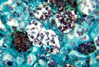
Analysis of the biopsy specimen identified the presence of diffuse inflammatory disease (a granulomatous disease with giant-cell formation), but no evidence of carcinoma. Histologically, a large number of yeast forms with some budding was seen on Gomori methenamine silver and periodic acid-Schiff staining. A test for acid-fast bacilli was negative. These findings were consistent with the presence of histoplasmosis. The lung lesion detected on x-ray was a calcified granuloma and was consistent with a histoplasmoma.
Upon further questioning, the patient provided a history of tongue and buccal ulcerations during the previous 4 years. These lesions had been treated with an antifungal agent prescribed by both a dentist and by an infectious disease specialist. The patient had lived in Connecticut his entire life, although he had spent 13 weeks in Missouri for military training approximately 20 years earlier.
We wanted to administer intravenous antifungal therapy, but the patient refused. Therefore, we prescribed a several-month course of an oral antifungal (itraconazole). Follow-up videostroboscopy during itraconazole treatment revealed a significant resolution of the laryngeal deformity (figure 2).
Histoplasma capsulatum is a nonencapsulated, budding fungus of the Moniliaceae family. Endemic to the Southeast, Midwest and central region of the United States, the fungus lives in moist soil. The fungus is acquired when patients breathe in the spores, which then settle in the lungs. In some cases, patients can progress to an acute disseminated state and exhibit upper-airway manifestations, including ulcers and erosion. Treatment is typically intravenous amphotericin B. (1, 2)
References
(1.) Reibel JF, Jahrsdoerfer RA, Johns MM, Cantrell RW. Histoplasmosis of the larynx. Otolaryngol Head Neck Surg 1982;90:740-3.
(2.) Sataloff RT, Wilbom A, Prestipino A, et al. Histoplasmosis of the larynx. Am J Otolaryngol 1993;14:199-205.
From the Division of Otolaryngology--Head and Neck Surgery, University of Connecticut, Farmington.
COPYRIGHT 2002 Medquest Communications, LLC
COPYRIGHT 2002 Gale Group


