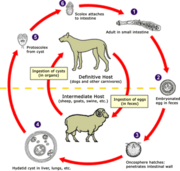
Echinococcus organisms taken from a hydatid cystEchinococcus life cycle (click to enlarge)
Find information on thousands of medical conditions and prescription drugs.

Hydatidosis

Echinococcosis, also known as hydatid disease, is a potentially fatal parasitic disease that can affect many animals, including wildlife, commercial livestock and humans. more...

Home
Diseases
A
B
C
D
E
F
G
H
Hairy cell leukemia
Hallermann Streiff syndrome
Hallux valgus
Hantavirosis
Hantavirus pulmonary...
HARD syndrome
Harlequin type ichthyosis
Harpaxophobia
Hartnup disease
Hashimoto's thyroiditis
Hearing impairment
Hearing loss
Heart block
Heavy metal poisoning
Heliophobia
HELLP syndrome
Helminthiasis
Hemangioendothelioma
Hemangioma
Hemangiopericytoma
Hemifacial microsomia
Hemiplegia
Hemoglobinopathy
Hemoglobinuria
Hemolytic-uremic syndrome
Hemophilia A
Hemophobia
Hemorrhagic fever
Hemothorax
Hepatic encephalopathy
Hepatitis
Hepatitis A
Hepatitis B
Hepatitis C
Hepatitis D
Hepatoblastoma
Hepatocellular carcinoma
Hepatorenal syndrome
Hereditary amyloidosis
Hereditary angioedema
Hereditary ataxia
Hereditary ceroid...
Hereditary coproporphyria
Hereditary elliptocytosis
Hereditary fructose...
Hereditary hemochromatosis
Hereditary hemorrhagic...
Hereditary...
Hereditary spastic...
Hereditary spherocytosis
Hermansky-Pudlak syndrome
Hermaphroditism
Herpangina
Herpes zoster
Herpes zoster oticus
Herpetophobia
Heterophobia
Hiccups
Hidradenitis suppurativa
HIDS
Hip dysplasia
Hirschsprung's disease
Histoplasmosis
Hodgkin lymphoma
Hodgkin's disease
Hodophobia
Holocarboxylase...
Holoprosencephaly
Homocystinuria
Horner's syndrome
Horseshoe kidney
Howell-Evans syndrome
Human parvovirus B19...
Hunter syndrome
Huntington's disease
Hurler syndrome
Hutchinson Gilford...
Hutchinson-Gilford syndrome
Hydatidiform mole
Hydatidosis
Hydranencephaly
Hydrocephalus
Hydronephrosis
Hydrophobia
Hydrops fetalis
Hymenolepiasis
Hyperaldosteronism
Hyperammonemia
Hyperandrogenism
Hyperbilirubinemia
Hypercalcemia
Hypercholesterolemia
Hyperchylomicronemia
Hypereosinophilic syndrome
Hyperhidrosis
Hyperimmunoglobinemia D...
Hyperkalemia
Hyperkalemic periodic...
Hyperlipoproteinemia
Hyperlipoproteinemia type I
Hyperlipoproteinemia type II
Hyperlipoproteinemia type...
Hyperlipoproteinemia type IV
Hyperlipoproteinemia type V
Hyperlysinemia
Hyperparathyroidism
Hyperprolactinemia
Hyperreflexia
Hypertension
Hypertensive retinopathy
Hyperthermia
Hyperthyroidism
Hypertrophic cardiomyopathy
Hypoaldosteronism
Hypocalcemia
Hypochondrogenesis
Hypochondroplasia
Hypoglycemia
Hypogonadism
Hypokalemia
Hypokalemic periodic...
Hypoparathyroidism
Hypophosphatasia
Hypopituitarism
Hypoplastic left heart...
Hypoprothrombinemia
Hypothalamic dysfunction
Hypothermia
Hypothyroidism
Hypoxia
I
J
K
L
M
N
O
P
Q
R
S
T
U
V
W
X
Y
Z
Medicines

The Echinococcosis Cycle

The disease results from infection by tapeworm larvae of the genus Echinococcus - notably E. granulosus, E. multilocularis, and E. vogeli.

Like many parasite infections, the course of Echinococcus infection is complex. The worm has a life cycle that requires definitive hosts and intermediate hosts. Definitive hosts are normally carnivores such as dogs, while intermediate hosts are usually herbivores such as sheep and cattle. Humans also function as intermediate hosts, although they are usually a 'dead end' for the parasitic infection cycle.

The disease cycle begins with an adult tapeworm infecting the intestinal tract of the definitive host. The adult tapeworm then produces eggs which are expelled in the host's feces.

Intermediate hosts become infected by ingesting the eggs of the parasite. Inside the intermediate host, the eggs hatch and release tiny hooked embryos which travel in the bloodstream, eventually lodging in an organ such as the liver, lungs and/or kidneys. There, they develop into hydatid cysts. Inside these cysts grow thousands of tapeworm larvae, the next stage in the life cycle of the parasite. When the intermediate host is predated or scavenged by the definitive host, the larvae are eaten and develop into adult tapeworms, and the infection cycle restarts.

Disease Symptoms

As already noted, Echinococcus infection causes large cysts to develop in intermediate hosts. Disease symptoms arise as the cysts grow bigger and start eroding and/or putting pressure on blood vessels and organs. Large cysts can also cause shock if they happen to rupture.

Infection with E. granulosus typically results in the formation of cysts in the liver, lungs, kidney and spleen of the intermediate host. In a full-blown infection, cysts can be larger than a soccer ball. This condition is also known as cystic hydatid disease and can sometimes be successfully treated with surgery to remove the cysts.

Infection with E. multilocularis results in the formation of dense parasitic tumors in the liver, lungs, brain, and other organs. This condition, also called alveolar hydatid disease is more likely to be fatal.

Unlike intermediate hosts, definitive hosts are usually not hurt very much by the infection. Sometimes, a lack of certain vitamins and minerals can be caused in the host by the very high demand of the parasite.

Disease Prophylaxis

There are several strategies to prevent Echinococcosis, most of which involve disruption of the parasite's life cycle. For instance, feeding raw offal to work dogs is a key point of infection in a farm environment and is strongly discouraged. Also, basic hygiene practices such as thoroughly cooking food and vigorous hand washing before meals can prevent the eggs entering the human digestive tract.

Read more at Wikipedia.org


[List your site here Free!]


15 Years In Surgical Management Of Pulmonary Hydatidosis - Abstract
From CHEST, 10/1/00 by Adel K Ayed

Adel K Ayed, FCCP(*) and Rafat Shalaby. Department of Surgery, Faculty of Medicine, Kuwait University, Kuwait and Chest Diseases Hospital, Kuwait.

PURPOSE: To evaluate the clinical presentation, diagnosis, management and outcome of patients with pulmonary hydatidosis.

METHODS: Retrospective review of 60 patients with hydatid disease of the lung and 15 years follow-up.

RESULTS: There were 35 males and 25 females with a mean age of 28.4 years. The most common presenting symptoms were cough (41 patients). Chest x-ray showed round show in 42 patients; 19 cases were of vigorous size [is greater than] 10cm, ill defined lung shadow in 8, pleural effusion in 6, cysts with air fluid level in 4 patients. Fifteen cases had cysts in other organs; 11 cases liver hydatid cyst, 2 in the spleen, and kidney and heart are one each. Preoperative diagnosis was pulmonary hydatid cyst in 41 patients. Different operative procedures were used to manage the hydatid cysts. The mean hospital stay was 9 days. The postoperative complications occurred in 9 patients; prolonged air leak in 4 patients, pleural effusion in 3, pneumothorax and wound infection in one patient each. One patient died in this series from pulmonary embolism. In follow-up period, 4 recurrences have been noted.

CONCLUSION: Surgical excision of pulmonary hydatid cysts with a maximum preservation of lung parenchyma is the mainstay of treatment.

COPYRIGHT 2000 American College of Chest Physicians
COPYRIGHT 2001 Gale Group

Return to Hydatidosis
Home Contact Resources Exchange Links ebay