Accepted June 10, 1999
Rapid enzyme linked immunosorbent assay (ELISA) was compared with the standard ELISA and indirect haemagglutination (IHA) techniques for the diagnosis of human hydatidosis. Eighty nine serum samples including 17 from hydatidosis patients (10 surgically confirmed and 7 clinically suspected), 50 from patients with other parasitic diseases and 22 samples from normal healthy individuals were analysed for anti-hydatid IgG antibodies using sheep hydatid cyst fluid antigen. The sensitivity and specificity respectively was found to be 82.3 and 100 per cent by rapid ELISA; 88.23 and 90.27 per cent by standard ELISA and 70.58 and 100 per cent by IHA technique. No cross reactions were observed with rapid ELISA technique using samples from cysticercosis and amoebiasis patients and normal healthy controls. The present study indicates that rapid ELISA can easily be performed in place of the standard ELISA for the serodiagnosis of human hydatidosis with the advantage of minimising reporting time and manpower hours.
Key words Diagnosis - enzyme linked immunosorbent assay - hydatidosis
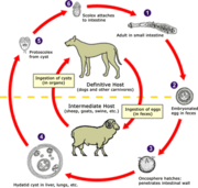
Human hydatidosis caused by the larval forms of Echinococcus granulosus, occurs world-wide due to the varied and yet cosmopolitan distribution of the intermediate hosts (sheep, pig, cattle etc.) and definitive host (dog)1. Though the liver is the most frequently involved site, the cysts can develop in almost all organs of the body 1-3.
The laboratory diagnosis mainly rests upon the detection of anti-hydatid antibodies in serum samples and usually supplements clinical and radiological data. Among the serological techniques, the enzyme linked immunosorbent assay (ELISA) is the most sensitive and specific technique using crude hydatid antigens4. Rapid diagnostic techniques with reliable results are required so as to provide a prompt diagnosis. Although commercially available kits offer this approach they are very costly and their use may not be cost effective in most laboratories. The present study was aimed at the assessment of the diagnostic accuracy of rapid ELISA in comparison to standard ELISA and IHA for the detection of anti-hydatid IgG antibodies in serum samples.
Material & Methods
Specimens: Sixty seven serum samples were obtained from patients attending the Nehru Hospital attached to the Postgraduate Institute of Medical Education and Research (PGIMER), Chandigarh. These comprised 17 samples from patients clinically suspected to have hydatidosis (10 from surgically confirmed and 7 from clinically suspected hydatid disease) and 50 from patients with other parasitic diseases (Table). Twenty two serum samples were also collected from normal healthy individuals after obtaining their consent and who were found negative for intestinal helminthic infestation by repeated stool examination.
Antigen: Antigen was prepared5 from the hydatid cysts obtained from naturally infected sheep, from the local slaughter house. Briefly, hydatid cyst fluid was drawn aseptically from the cysts and 5 mM phenylmethylsulphonyl fluoride was added. It was then centrifuged at 3000 g for 20 min to settle the protoscolices and the supernatant was taken as antigen and stored at -20 deg C till further use. The antigen protein concentration was estimated by the method of Lowry et al with bovine serum albumin as a reference standard. Standard ELISA : The ELISA was performed essentially by the method of Voller et aP with slight modifications. Using checker board titration, antigen, serum and antihuman IgG HRP conjugate (Dakopatts, Denmark) dilutions were found to be 5 (mu)g/well; 1:1280 and 1:1000 respectively. The only modification made in the standard ELISA procedure7 was that dilutions of serum samples were carried out in PBS-T buffer with 1 per cent BSA and the A^sub 492^ of the enzymatic product was determined directly in the wells with an automated ELISA reader (A4 Eurogenetics, Tessenderle, Belgium). Positive and negative serum samples were included in each plate along with the substrate and buffer blanks.
Rapid ELISA : The technique used was basically the same as that for standard ELISA, with similar optimum dilutions of antigen and conjugate, except that the plates were coated with antigen by incubation at 4 deg C overnight or at 37 deg C for 1h8 and further all the incubation periods at subsequent steps were reduced from one hour to 15 min each.
Indirect haemagglutination assay: The IHA test was carried out as detailed earlier9. Briefly, the test was done by the use of tanned sheep erythrocytes sensitized with hydatid fluid antigen. The working dilutions of antigen and serum samples were predetermined by checker board titration method. Titer >= 1:80 was considered positive.
Results & Discussion
The cyst localization was found to be hepatic in 15 and multiple organ involvement in 2 of the 17 patients clinically suspected to have hydatidosis.
The rapid ELISA gave a cut-off OD value of 0.910 and standard ELISA 0.686 (mean +/- 3SD of 22 serum samples from normal healthy individuals). Therefore, serum samples with an OD value >= 0.910 and >= 0.686 were considered positive for anti-hydatid antibodies by rapid and standard ELISA respectively. All the 10 samples from surgically confirmed hydatidosis and 4 of 7 clinically suspected patients had positive antibody response while none of the samples from cysticercosis and amoebiasis patients and normal healthy individuals were positive by rapid ELISA technique. The sensitivity and specificity by rapid ELISA was 82.3 and 100 per cent respectively. All 10 samples from surgically confirmed and 5 of 7 clinically suspected hydatidosis patients and 7 of 34 cysticercosis patients were found positive by the standard ELISA. None of the samples from amoebiasis patients and healthy individuals were reactive. The sensitivity and specificity by standard ELISA was assessed as 88.23 and 90.27 per cent respectively. Positive antibody response by IHA technique was found in all the 10 samples from surgically confirmed and in-2 of 7 clinically suspected patients of hydatidosis. All samples from healthy individuals were found negative while samples from cysticercosis and amoebiasis were not tested by IHA technique (Table). Although the sensitivity of this technique was found to be 70.5 per cent, yet specificity was 100 per cent, when evaluated in samples from normal healthy individuals. Samples from 2 hydatidosis patients with multiple organ involvement were positive by all the three techniques.
Detection of anti-hydatid antibodies helps in the diagnosis of human hydatidosis. ELISA has been reported to be highly sensitive and specific technique for the diagnosis of various parasitic diseases, I'. The use of rapid ELISA technique has the potential to reduce the overall time from specimen collection to final identification, thereby, minimising the reporting time and manpower hours. Commercial kits with the use of rapid ELISA have become available and have been demonstrated to be highly reliable for the diagnosis of parasitic diseases12. However, due to high cost of imported kits, it may not be economically feasible to use this approach on large number of samples, in endemic places, for routine diagnosis.
Hydatid cyst fluid (HCF) antigen has been found to be a good source of antigen for the serodiagnosis of hydatid disease4. Use of purified antigenic fraction/s in highly sensitive techniques has obviated cross reactions with most of the other infections 13,14, yet it may sometimes pose problems5,15. In the present study, the rapid ELISA, standard ELISA and IHA techniques were found equally sensitive (100%) for the detection of antihydatid antibodies to crude HCF antigen in surgically confirmed patients of hydatidosis. However, due to lack of follow up in the 7 clinically suspected patients, it is difficult to ascertain whether these patients had hydatidosis or any other disease. However, with rapid ELISA no cross reactions were noticed with samples from cysticercosis or amoebiasis patients, thereby indicating the high specificity of the rapid technique.
In conclusion, results of the study indicate that rapid ELISA technique can be used for the serodiagnosis of human hydatidosis. It is a rapid, simple, valuable and convenient technique which may serve as an useful adjunct to the clinical and radiological diagnosis.
Acknowledgment The financial assistance provided by the Indian Council iff Medical Research, New Delhi, is acknowledged.
References
Beaver PC, Jung RC. Genus Echinococcus in clinical parasitology, 9th ed. Philadelphia: Lea & Febiger; 1984 p. 526-38.
2. Sharma A, Abraham J. Multiple giant hydatid cysts of the brain. Case report. J Neurosurg 1982; 57: 413-5. 3. Wani NA. Hydatid disease of the spleen. Indian J Surg 1993:
55: 155-60.
4. Wattal C, Malla N, Khan IA, Agarwal SC. Comparative evaluation of enzyme linked immunosorbent assay for the diagnosis of pulmonary echinococcosis. J Clin Microbiol 1986; 24: 41-6.
S. Hernandez A, Nieto A. Induction of protective immunity against murine secondary hydatidosis. Parasite Immunol 1994; 16: 537-44.
6. Lowry OH, Rosebrough NJ, Farr AL, Randall RJ. Protein measurement with the folin phenol reagent. JBiol Chem 1951; 193: 265-75.
7. Voller A, Bidwell DE, Bartlett A. Enzyme immunoassays in diagnostic medicine - Theory and practice. Bull World Health Organ 1976; 53: 55-65.
8. Mohammad IN, Heiner DC, Miller BL, Goldberg MA, Kagan IG. Enzyme linked immunosorbent assay for the diagnosis of cerebral cysticercosis. J Clin Microbiol 1984; 20: 775-9. 9. Mahajan RC, Ganguly NK, Chitkara NL. Evaluation of immunodiagnostic techniques for hydatid disease. Indian J Med Res 1976; 64: 405-9.
10. Pahuchonma W, Janitschke K. Serodiagnosis of parastic infections by ELISA with different antigens. Southeast Asian J Trop Med Public Health 1987; IB: 193-6.
1I. Maddison SE. Serodiagnosis of parasitic diseases. Clin Microbiol Rev 1991; 4: 457-69.
12. Kraoul L, Adjmi H, Lavarde V, Pays JF, Tourte-Schaefer C, Hennequin C. Evaluation of a rapid enzyme immunoassay for diagnosis of hepatic amoebiasis. J Clin Microbiol 1997; 35: 1530-2.
13. Maddison SE, Slemenda SB, Schantz PM, Fried JA, Wilson M. Tsang VC. A specific diagnostic antigen of Echinococcus
granulosus with an apparent molecular weight of 8 kDA. Am J Trop Med Hyg 1989; 40: 377-83.
14. Kanwar JR, Kaushik SP, Sawhney IMS, Kamboj MS, Mehta SK, Vinayak VK. Specific antibodies in serum of patients with hydatidosis recognized by immunoblotting. J Med Microbiol 1992; 36: 46-51.
15. Kharebov A, Nahmias J, El-On J. Cellular and humoral immune responses of hydatidosis patients to Echinococcus granulosus purified antigens. Am J Trop Med Hyg 1997: 57: 619-25.
Reprint requests: Dr Nancy Malla, Head, Department of Parasitology, Postgraduate Institute of Medical Education & Research, Chandigarh 160012
Manjit Kaur, R.C. Mahahan & Nancy Malla
Department of Parasitology, Postgraduate Institute of Medical Education & Research, Chandigarh
Copyright Indian Council of Medical Research Jul 1999
Provided by ProQuest Information and Learning Company. All rights Reserved


