A 37-year-old Greek woman presented to her physician's office for a routine physical examination. She did not complain of any symptoms or active medical problems. Her medical history was notable only for mitral valve prolapse. She did not smoke, drink alcohol, or use illicit drugs. Physical examination revealed a II/VI systolic murmur over the mitral area. Deep palpation of the right upper abdominal quadrant caused mild discomfort and pain. Results of the rest of the physical examination were normal.
The patient underwent laboratory and imaging testing to evaluate tenderness of the right upper abdominal quadrant that was noted during physical examination. The laboratory testing, which included complete blood count and measurement of serum glucose, creatinine, total lipid, cholesterol, triglyceride, alanine transaminase, aspartate transaminase, gamma glutamyl trans-peptidase, and alkaline phosphatase levels, did not reveal any abnormal findings. The erythrocyte sedimentation rate was elevated slightly (26 mm in the first hour [normal values less than 20 mm]). Ultrasonography of the right upper quadrant revealed a well-circumscribed mass with mixed echographic pattern in the right hepatic lobe. Contrast-enhanced computed tomography (CT) of the abdomen was performed subsequently (Figures 1 and 2).
[FIGURES 1-2 OMITTED]
Question
Based on the patient's history, physical examination, and CT findings, which one of the following is the correct diagnosis?
[] A. Hepatocellular carcinoma.
[] B. Pyogenic abscess.
[] C. Simple cyst.
[] D. Echinococcal cyst.
[] E. Metastatic carcinoma.
Discussion
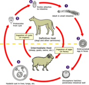
The answer is D: echinococcal cyst. Solitary or multiple cystic lesions of the liver may be caused by a variety of conditions, including primary and metastatic neoplasms, non-neo-plastic cysts, and infections. (1) Echinococciasis (hydatid cyst) is a parasitic disease most often caused by Echinococcus granulosus. Dogs are the host animal, passing eggs in their feces. Echinococciasis always should be included in the differential diagnosis of symptomatic or asymptomatic right upper quadrant masses, particularly in patients who are from or have traveled to endemic areas. Extrahepatic sites include the bones, brain, and lungs.
Ultrasonography and CT imaging are the cornerstone for the diagnosis, classification of the stage of progression, and determination of the best mode of treatment. Blood tests are available, although the limitations of serum testing should be taken into consideration: the sensitivity of serum echinococcal antigens ranges from 80 to 95 percent for hepatic-limited echinococcal disease, and is lower for solitary extrahepatic echinococcal cysts. (2,3) Specificity ranges from 88 to 96 per-cent. (3) Needle biopsy and cyst aspiration are options, but often are avoided because of the risk of cyst rupture.
The CT imaging in this patient is not typical of echinococciasis. Echinococcal cyst can lead to peculiar findings on different imaging techniques, depending on which parts of the cyst are captured in the image and on the stage of disease. Although CT sections of the lower and central part of the lesion exhibited the typical well-demarcated, hypodense cyst (Figure 1), (4) CT sections of the upper part of the lesion exhibited hyperdense, separated, peripheral areas (Figure 2). They may represent calcified daughter hydatid cysts (5) separated by fibrous tissue or, more likely, a redundant, folded, inner germinal wall of a cyst. (1)
Echinococciasis continues to be endemic in Greece, although its frequency has decreased over the past two decades. Areas with high endemicity include the Mediterranean basin, the Middle East, Central Asia, Chile, and Argentina. (6,7)
Serum echinococcal antigens were negative in this patient, but biopsy of the excised specimen confirmed the diagnosis of echinococciasis. Treatment included surgical excision of the mass, with 400 mg of albendazole per day for one week before and two weeks after surgery.
Hepatocellular carcinoma is unlikely because of the imaging characteristics of the liver lesion. Usually, such lesions would have irregular borders. In most patients, hepatocellular carcinoma presents after the fifth decade of life, and is related to a history of chronic liver disease (e.g., viral hepatitis, cirrhosis).
Pyogenic liver abscess usually manifests with fever. Chills, rigors, or malaise also may be present, and some patients appear to be septic.
A simple cyst of the liver does not have calcified areas. In addition, simple cysts do not have mixed echogenic characteristics unless they are infected or hemorrhagic.
Liver metastases frequently present with multiple hepatic lesions on diagnosis. Usually the patient already has symptoms and/or signs from the primary cancer.
REFERENCES
(1.) Baden LR, Elliott DD. Case records of the Massachusetts General Hospital. Weekly clinicopathological exercises. Case 4-2003. A 42-year-old woman with cough, fever, and abnormalities on thoracoabdominal computed tomography. N Engl J Med 2003;348:447-55.
(2.) Gottstein B, Reichen J. Echinococciasis/hydatidosis. In: Manson P, Cook GC, Zumla AI, eds. Manson's Tropical diseases. 21st ed. London: Saunders, 2003:1564-5.
(3.) King CH. Echinococcosis (hydatid and alveolar cyst disease). In: Mandell GL, Douglas RG, Bennett JE, Dolin R, eds. Mandell, Douglas, and Bennett's Principles and practice of infectious diseases. 4th ed. New York: Churchill Livingstone, 1995:2550-1.
(4.) Tuzun M, Hekimoglu B. Pictorial essay. Various locations of cystic and alveolar hydatid disease: CT appearances. J Comput Assist Tomogr 2001;25:81-7.
(5.) Sayek I, Onat D. Diagnosis and treatment of uncomplicated hydatid cyst of the liver. World J Surg 2001;25: 21-7.
(6.) McManus DP, Zhang W, Li J, Bartley PB. Echinococcosis. Lancet 2003;362:1295-304.
(7.) Suwan Z. Sonographic findings in hydatid disease of the liver: comparison with other imaging methods. Ann Trop Med Parasitol 1995;89:261-9.
IOANNIS A. BLIZIOTIS, M.D.
SOFIA S. KASIAKOU, M.D.
MARIA BALOYANNI, M.D.
MATTHEW E. FALAGAS, M.D., M.SC.
Alfa Institute of Biomedical Sciences (AIBS)
and Henry Dunant Hospital Athens, Greece
E-mail: matthew.falagas@tufts.edu
COPYRIGHT 2005 American Academy of Family Physicians
COPYRIGHT 2005 Gale Group


