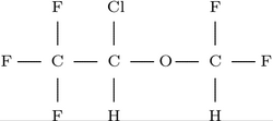
Two adults and two children with life-threatening asthma refractory to maximal standard therapy were treated with the inhalational anesthetic agent isoflurane. In each case, the temporal response to the initiation of therapy was striking. All patients survived and none experienced adverse reactions attributable to the drug. Rapid therapeutic benefit, minimal side effects, absence of cumulative toxicity, and ease of administration are factors supporting the use of isoflurane for patients with severe asthma.
ABG = arterial blood gas; PIP = peak inspiratory pressure
Inhalational anesthetic agents have been sporadically used for status asthmaticus unresponsive to maximal standard therapy.[1-5] Unfortunately, many agents have serious side effects and the delivery systems have been difficult to use outside the operating room, particularly in patients with high airway pressures.
Isoflurane, a halogenated ether, is an anesthetic agent that produces bronchodilation through a number of mechanisms.[6] Recent technologic developments allow isoflurane to be easily and safely administered for prolonged periods of time to patients with severe bronchospasm.
We describe its use in two adults and two children who were refractory to conventional therapy. All responded favorably to isoflurane over 16 to 48 hours, and none experienced significant side effects.
Case Reports
Case 1
A 20-year-old woman with a seven-year history of asthma was admitted to the hospital with a history of worsening bronchospasm for one week following a flu-like illness. She had been using albuterol, ipratropium, and beclomethasone inhalers without improvement. owing to increasing respiratory distress. On admission to the ICU, her heart rate (HR) was 120 beats per minute, respiratory rate (RR) was 18 breaths per minute, and blood pressure (BP) was 150/80 mm Hg. Initial arterial blood gas (ABG) determinations were a pH of 7.30, [Pco.sub.2] of 35 mm Hg, and [Po.sub.2] of 45 mm Hg. Complete blood cell count (CBC), electrolytes and liver function test results were normal.
Supplemental oxygen was given and an aminophylline infusion at 60 mg/h was begun. Albuterol 2.5 mg every four was alternated with altropine inhalations 1.2 every four hours, such that an inhalation treatment was received every two hours. Methylprednisolone 250 mg intravenously, (IV) was given every six hours. Theophylline levels were maintained within the therapeutic range. The inhaled bronchodilators were administered with increasing frequency due to worsening bronchospasm, until administration occurred every 15 minutes. Twelve hours after ICU admission, ABG values were a pH of 7.22, [Pco.sub.2] of 55 mm Hg, and [Po.sub.2] of 137 mm Hg. She was intubated and ventilated, after sedation with diazepam and paralysis with pancuronium bromide. Her bronchospasm worsened and ventilation became increasingly, difficult. Despite manipulations in inspiratory flow, tidal volume, and RR, [Pco.sub.2] rose to mm Hg, and peak inspiratory pressure (PIP) was in excess of 60 cm [H.sub.2]O.
Isoflurane inhalation was initiated, using a ventilator (Siemens Servo 900C) with attached vaporizer (Siemens Electronics, Calgary, Canada). Treatment with all other medications except steroids was discontinued. The isoflurane concentration was quickly increased to 1 percent. Over the next 90 minutes, PIP fell to 50 cm [H.sub.2]O and static compliance rose from 0.012 to 0.02 L/cm [H.sub.2]O. The Pa[CO.sub.2] fell to 44 mm Hg and pH rose to 732. Within five hours there was marked clinical improvement. Table I shows the change in dynamic compliance, [Pco.sub.2], and HR during the use of isoflurane. The mean arterial BP remained above 70 mm Hg throughout therapy.
Isoflurane was continued for 16 hours and therapy with albuterol and aminophylline recommenced. Isoflurane treatment was discontinued when bronchospasm resolved and it seemed likely that the patient would tolerate extubation. Awakening occurred in 20 minutes, and she was extubated 40 minutes after the isoflurane treatment was discontinued.
The remainder of her hospital course was unremarkable. Although her aspartate aminotransferase (AST) and lactate dehydrogenase (LDH) levels increased transiently on day 2 to 170 U/L and 412 U/L, respectively, the values returned to normal the next day. There was no clinical evidence of hepatic damage. All other laboratory values remained within normal limits.
Case 2
An 18-year-old male patient, with a long history of severe asthma since childhood, awoke at home with severe dyspnea. Long-term medications included theophylline, cromolyn sodium (cromoglycate sodium), and albuterol. He arrived in the Emergency Department unconscious and had gasping respirations. Initial ABG values were a pH of 6.77, [Pco.sub.2] of 162 mm Hg, and [Po.sub.2] of 133 mm Hg. After intubation he was given aminophylline 150 mg IV loading dose, followed by an infusion at 40 mg/h, methylprednisolone 250 mg every six hours IV, albuterol 20 [mu]min IV, and ipratropium 0.5 mg by inhalation, alternating with albuterol 5 mg by inhalation every 15 minutes. Fentanyl, diazepam, and pancuronium bromide were given as needed.
One bour after intubation, ABG values were a pH of 6.85, [Pco.sub.2] of 145 mm Hg, and [Po.sub.2] of 499 mm Hg ([FIo.sub.2] of 1.0). The PIP was 72 cm [H.sub.2]O. Treatment with 1 percent isoflurane was commenced. Within one hour, [Pco.sub.2] fell to 71 mm Hg and the isoflurane concentration was decreased to 0.6 percent. Because the PIP remained at 70 cm [H.sub.2]O, bronchoscopy was performed for the removal of mucous plugs, but few were found. He was maintained on a regimen of aminophylline, methylprednisolone, and inhaled albuterol while ventilated. After 34 hours of isoflurane anesthesia, and once he had been stable for several hours with a [Pco.sub.2] of 35 mm Hg and a PIP of 36 cm [H.sub.2]O, the isoflurane treatment was discontinued. He was placed on a T-piece 55 minutes after the isoflurane treatment was stopped and was extubated four hours later. He had an uneventful hospital course. No hepatic or renal biochemical abnormalities ensued.
Case 3
A 27 month-old-boy was admitted to the pediatric ICU for the third time suffering from an exacerbation of asthma. The day prior to hospital admission he had been exposed to grass and pollen on a farm. At a rural hospital he was given albuterol, prednisone, and oral theophylline and transferred to our hospital. En route, he received hydrocortisone 125 mg IV and albuterol inhalations. The ABG values on admission were a pH of 6.94, [Pco.sub.2] of 132 mm Hg, and [Po.sub.2] of 79 mm Hg. Heart rate was 168 beats per minute and RR was 22 breaths per minute. Chest roentgenogram showed marked hyperinflation with no consolidation or pneumothorax. Following sedation with fentanyl and paralysis with pancuronium bromide, he was intubated. Subsequent ABG values were pH of 7.01, [Pco.sub.2] of 122 mm Hg, and [Po.sub.2] of 106 mm Hg (FIo.sub.2] of 0.7). The PIP was 44 cm [H.sub.2]O. Positive end-expiratory pressure of 4 cm [H.sub.2]O was added. Drug therapy included methylprednisolone 15 mg IV every eight hours, albuterol 2 [mu]g/kg/min IV, atropine 0.6-mg inhalations, and aminophylline 10 mg/h IV (theophylline levels maintained at 16 to 20 mg/L).
The following day, he developed a pneumothorax and underwent chest tube thoracostomy. The PIP was 74 cm [H.sub.2]O, and ABG values revealed a pH of 7.22, [Pco.sub.2] of 72 mm Hg, and [Po.sub.2] of 90 mm Hg. Isoflurane treatment was initiated and rapidly increased to 2.2 percent. Other inhalational agents were discontinued. Within an hour, inspiratory pressures decreased to 62 cm [Hsub.2]O and ABG values were pH of 7.26, [Pco.sub.2] of 66 mm Hg, and [Po.sub.2] of 106 mm Hg. Over the next 18 hours, PIP fell to 46 cm [H.sub.2]O. The isoflurane concentration was slowly decreased and discontinued 30 hours after initiation, at a time when the PIP was 40 cm [H.sub.2]O and the [Pco.sub.2] was 48 mm Hg. The patient remained paralyzed, but eight hours later, the child was opening his eyes as the effects of pancuronium dissipated. The child remained intubated for another 3 1/2 days while receiving standard conventional therapy of steroids, [beta]-adrenergic agonists, and theophylline compounds.
Case 4
A six-year-old girl with life-long asthma was admitted to a rural hospital. During the prior two years, she had been admitted to the hospital with exacerbations of asthma every one to two months. Treatment at the rural hospital included theophylline 550 mg/day in divided doses, albuterol, and cromolyn sodium inhalations. Therapy was intensified by adding fenoterol inhalations every hour and administering hydrocortisone 100 mg IV.
On arrival in the pediatric ICU, she was in severe respiratory distress. Respiratory was 50 breaths per minute, BP was 106/75 mm Hg, and HR was 160 beats per minute. Chest roentgenogram showed hyperinflation without evidence of consolidation. Subsequently, she developed left upper lobe collapse and left-sided pneumothorax that was treated with chest tube thoracostomy,.
She was begun on a regimen of aminophylline infusion 20 mg/h, maintaining a theophylline level at 10 mg/L, albuterol 2.5 mg inhalation hourly, and atropine 0.2-mg inhalations every two hours. Hydrocortisone was continued at 100 mg IV every six hours. Despite this her condition deteriorated, and ABG values showed a pH of 7.23, [Pco.sub.2] of 50 mm Hg, and [Po.sub.2] of 51 mm Hg. The RR was 40 beats per minute. Consequently, she was sedated with Fentanyl and paralyzed with pancuronium bromide for intubation and mechanical ventilation. Cefotaxime 500 mg IV every six hours was initiated because of a temperature of 38 Degree C, white blood cell count of 20,000/cu mm, and elevated bands of 17 percent. Pulmonary toilet and saline solution instillations performed every two hours resulted in removal of mucous.
Despite this, there were high PIPs of 56 to 60 cm [H.sub.2.O] and ABG values showed a pH of 7.07, [Pco.sub.2] of 60 mm Hg, and [Po.sub.2] of 84 mm Hg ([FIo.sub.2] of 0.75). She was commenced on 1 percent isoflurane therapy that was increased to 2.5 percent over one hour. After nine hours of therapy, ABG values were pH of 7.38, [Pco.sub.2] of 45 mm Hg, and [Po.sub.2] of 90 mm Hg ([FIo.sub.2] of 0.95), while the PIPs had decreased to 50 cm [H.sub.2.O]. Isoflurane therapy was maintained for 38 hours. It was discontinued when the PIP reached 40 cm [H.sub.2.O], at a time when the [Pco.sub.2] was 49 mm Hg. Seven hours after the isoflurane therapy was discontinued, the child was moving spontaneously and by ten hours, she was awake and alert. She was discharged from the hospital eight days later. There was no evidence of hepatic or renal toxicity.
DISCUSSION
Isoflurane produces bronchodilation through [beta]-adrenergic receptor stimulation, direct relaxation of bronchial smooth muscle, antagonism of the action of acetylcholine and histamine, and interference with hypocapnic bronchoconstriction.[6] Thus, a patient who is already receiving maximum doses of standard bronchodilators may show an additional response. As our case reports suggest, isoflurane acts rapidly and may be lifesaving while high-dose corticosteroids take effect. In contrast, ketamine, an intravenous anesthetic agent that has also been used in asthma, acts by adrenergic stimulation. Little response is seen in patients receiving large doses of beta-agonists and theophylline.[7]
There are several advantages of isoflurane over other inhalational anesthetic agents. Historically, diethyl ether and cyclopropane were used, but their extreme flammability precluded their use in electrically active environments. Isoflurane is the least fat soluble of the anesthetic vapors and has the lowest blood gas solubility coefficient.[8] Consequently, depth of anesthesia can be most rapidiy adjusted with isoflurane, and time to recovery of consciousness is short, despite prolonged use.[9,10]
All halogenated vapors are proarrhythmic, especially in the presence of adrenergic stimulants. Nevertheless, isoflurane is the least likely to cause arrhythmias. In dogs, at least four times the dose of epinephrine is needed to produce ventricular extrasystoles in the presence of isoflurane as compared with halothane (in equi-anesthetic doses).[11]
Halogenated hydrocarbons have been implicated in severe hepatic and renal toxicity. While halothane and enflurane are not directly toxic, their metabolic products appear to be responsible for a rare form of hepatic necrosis. Enflurane is also responsible for fluoride-induced renal failure. Isoflurane undergoes minimal metabolism (< O.2 percent),[8] and to our knowledge, there are no documented cases of isoflurane-induced hepatic or renal injury.
Isoflurane causes dose-dependent hypotension by peripheral vasodilation. Cardiac output is unchanged, while heart rate increases by 5 to 20 percent as the drug concentration is increased to 2 percent.[8] Hypotension is usually responsive to plasma volume expansion, but occasionally vasopressors are needed. All patients in our series responded to volume expansion.
Isoflurane causes amnesia in low concentrations and general anesthesia as the concentration is increased. Above 1.5 percent, other sedatives and muscle relaxants are rarely needed. Narcotics ought to be given for painful procedures, since isoflurane is a poor analgesic. if long-acting sedatives are avoided, as in our patients, awakening is rapid when isoflurane treatment is discontinued.
Until recently, it was very difficult to administer isoflurane in the ICU. Standard anesthetic ventilators do not achieve sufficiently high inspiratory pressures, nor do they have the necessary alarms and safety features for prolonged use outside the operating room. A standard ICU ventilator, (Siemens 900C), can be fitted with an isoflurane vaporizer. This ventilator generates inspiratory pressures up to 120 cm [H.sub.2.O] and provides essential alarms.
Scavenging exhaust gases in the ICU is necessary for preventing contamination with waste gases that can cause sedation. There is also concern about the effects of long-term anesthetic exposure for health care personnel.
We installed two waste gas scavenging systems into the operating room. This was simple and inexpensive, as the ICU is contiguous with the operating room.
An adequate scavenging device can be made by connecting the exhaust port of the ventilator to a T-piece that is attached to a 3-L reservoir bag. Wall suction is applied to the remaining limb of the T-piece until the reservoir bag partially fills with each breath, but never fully collapses. Leaks are readily detectable, since isoflurane has a characteristic pungent odor.
Since this is a nonrebreathing system, a great deal of isoflurane is vaporized hourly. The cost for 24 h of continuous use is high, but it is partially offset by decreased use of other sedatives and muscle relaxants.
Most authors have suggested that an anesthesiologist[1-5,12] should be in constant attendance of the patient, since isoflurane is unfamiliar to most critical care personnel. This is extremely difficult to organize, particularly if the drug is used for prolonged periods. We have dealt with this problem by providing educational material to the nurses in the ICU, supplemented bv bedside educational sessions for the nurse caring for the patient. An anesthesiologist (R.G.J.) initiated therapy with the drug and titrated the dose by increments of 0.5 percent until the PIP decreased, the BP fell, or a concentration of 2.5 percent was reached. If the BP fell, saline solution or albumin was infused until the BP increased to an acceptable level. The isoflurane concentration was then increased again at 0.5 percent increments. Once the patient was in stable condition while receiving a given concentration for one hour, the anesthesiologist was no longer in constant attendance. The nursing supervisors, who received more in-depth instruction, and who were experienced ICU nurses, were allowed to vary the concentration by increments of 0.5 percent. When the PIP began to decline, the isoflurane concentration was kept constant for several hours. Attempts were then made to decrease the concentration until reaching the minimal concentration that resulted in lower airway pressures, clinical improvement, and improved ventilation. The nurses were instructed to immediately discontinue the isoflurane treatment if the patients became severely hypotensive or if arrhythmias occurred. This circumstance never arose. During maintenance with isoflurane, narcotics were given for painful procedures and muscle relaxants were used if the patients coughed or became asynchronous with the ventilator. The nurse was in constant attendance at the bedside to guard against accidental ventilator disconnection or loss of airway control.
When PIP was less than 40 cm [H.sub.2.O] for several hours, and clinical assessment revealed marked improvement, isoflurane treatment was discontinued while an intensivist was present. Awakening was surprisingly rapid, occurring in 20 to 60 minutes. This is predicted theoretically[10] and confirmed in one other case report.[9] If bronchospasm worsened during awakening, the isoflurane dose was increased to its former level with rapid relief.
In a patient who is unresponsive to maximal conventional therapy for status asthmaticus, isoflurane may be the drug of choice for additional therapy. Its relatively mild side effects, lack of toxicity, sedative action, and ease of administration are significant advantages. Further clinical trials would seem to be indicated.
REFERENCES
[1] Meyer NE, Schotz S. Relief of severe intractable bronchial asthma with cyclopropane anesthesia: report of Case. J Allergy 1939; 10:239-40 [2] Schwartz SH. Treatment of status asthmaticus with halothane. JAMA 1984; 251:2688-89 [3] Bierman MI, Brown M, Muren O, Keenan RL, Glauser FL. Prolonged isoflurane anesthesia in status asthmaticus. Crit Care Med 1986; 14;9:832-33 [4] Parnass SM, Feld JM, Chamberlin WH, Segil LJ. Status asthmaticus treated with isoflurane and enflurane. Anesth Analg 1987; 66:193-95 [5] Revell S, Greenhalgh D, Absalom SR, Soni N. Isoflurane in the treatment of asthma. Anesthesia 1988; 43:477-79 [6] Hirshman CA, Edelstein G, Peetz S, Wayne R, Downes H. Mechanism of action of inhalational anesthesia on airways. Anesthesiology 1982; 56:107-11 [7] White PF, Way WL, Trevor AJ. Ketamine -- its pharmacology and therapeutic uses. Anesthesiology 1982; 56:119 [8] Eger EI. Isoflurane: a review. Anesthesiology 1981; 55:559-76 [9] Pearson J. Prolonged anesthesia with isoflurane. Anesth Analg 1985; 64:92-3 [10] Lowe JH, Ernst EA. The quantitative practice of anesthesia. Baltimore, Md: Williams & Wilkins; 1981 [11] Joas TA, Stevens WC. Comparison of the arrhythmic doses of epinephrine during forane, halothane and fluroxane anesthesia in dogs. Anesthesiology 1971; 35:48-53 [12] Kofke WA, Snider MT, Young RSK, Ramer JC. Prolonged low flow isoflurane anesthesia for status epilepticus. Anesthesiology 1985; 62:653-56
COPYRIGHT 1990 American College of Chest Physicians
COPYRIGHT 2004 Gale Group


