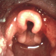
Study objective: To evaluate the practice of using nasopharyngoscopy without routine fiberoptic bronchoscopy for children presenting to a pediatric pulmonary practice with nonspecific noisy breathing.
Design: Retrospective chart review. Records of patients who underwent nasopharyngoscopy between January 1, 1990, and December 31, 1999, were reviewed. Follow-up was obtained by office records and direct contact with the patient's family and/or primary care physician.
Setting: Academic, tertiary care facility.
Results: Eighty-one children who underwent upper airway endoscopy to evaluate noisy breathing consistent with extrathoracic lesions were identified. One child had two evaluations separated by years for differing complaints, making a total of 82 procedures. Strider was the chief complaint in three fourths of the children. Half of the children with strider were found to have laryngomalacia. Long-term follow-up was available for 75 of 81 children, with median follow-up of 6 years (range, 1 to 13 years). No medical problems related to missed airway lesions developed in any infants initially evaluated using nasopharyngoscopy.
Conclusions: Nasopharyngoscopy without lower airway endoscopy can be used safely for the initial evaluation of noisy breathing in infants and children provided excellent follow-up is available.
Key words: bronchoscopy; laryngomalacia; pediatrics; strider; upper airway
Abbreviations: FOB = fiberoptic bronchoscopy; GERD = gastroesphageal reflux disease
**********
Infants and children who present to primary care physicians with the nonspecific complaint of noisy breathing often are referred to pediatric pulmonologists or otolaryngologists. Some have argued that the lower airway should be visualized in all children with hoarseness, stridor, and other upper airway sounds due to the possibility of finding a concomitant sublaryngeal lesion. (1-5) Since it is not obvious from the published material that clinically significant lesions would be missed, the practice at our institution has been to use procedures less invasive than full endoscopic visualization of the lower airways for the initial evaluation of children when the history and physical examination are highly suggestive of a sole upper airway problem. We reviewed the outcomes of our patients over a 10-year period to determine if our practice pattern was appropriate.
MATERIALS AND METHODS
Eighty-one children had nasopharyngoscopy performed between Jannuary 1, 1990, and December 31, 1999, lot evaluation of stridor, hoarseness, nonspecific "noisy breathing," and other suspected extrathoracic airway problems. Chart review and patient contact were approved by the Committee tier the Protection of Human Subjects in Research at the University of Massachusetts Medical School. Approximately one half of the patients have had continuing care in our clinic. Telephone calls were placed to families no longer seen in our clinic. In 16 cases, no contact could be made with the family. For 10 of these children, follow-up information was obtained from primary care providers. Six patients were unavailable for follow-up. Queries included the child's current respiratory status, whether an additional opinion regarding the airway problem had been sought at another institution, and whether further therapy beyond that recommended following our completed evaluation was necessary. Data collection was enhanced by canvassing physicians in attendance at a weekly meeting of the Massachusetts Pediatric Pulmonary Consortium and at quarterly meetings of the Massachusetts Cystic Fibrosis Newborn Screening Consortium. The core group of pediatric otolaryngologists from a major tertiary care airway clinic attend the weekly Pediatric Pulmonary Consortium.
Nasopharyngoscopy was performed at the discretion of the attending pulmonologist based on history and physical examination findings as outlined in Figure 1. As this was a retrospective chart review, there were no a priori conditions for performing nasopharyngoscopy. In general, children, with very, minimal stridor who were thriving, children with a history strongly suggestive of foreign body aspiration, and children with symptoms of lower airway lesions (homophonous wheezes) did not have nasopharyngoscopy performed. Nasopharyngoscopy was performed on children with stridor audible at a distance, retractions, poor growth, weak or hoarse cry, or symptoms worsening over time. In addition, some children with respiratory sounds suggestive of a fixed airway lesion had nasopharyngoscopy performed to role out an upper airway lesion.
[FIGURE 1 OMITTED]
A 3.3-mm flexible nasopharyngoscope was used (Model 3L; Machida; Orangeburg, NY) was used. The naris was anesthetized with 1% lidocaine jelly, and the scope was passed through the nose while the patient was in the supine position. Pulse oximeters and cardiac monitors were not routinely used. Full airway endoscopy or radiographic studies of the airway (including barium swallow) were obtained if nasopharyngoscopy was unrevealing or equivocal, or if the endoscopist was concerned about the possibility of a sublaryngeal lesion.
RESULTS
There were 82 nasopharyngoscopy procedures on 81 patients available for evaluation. During the time encompassed by this chart review, our center was staffed by two pediatric pulmonologists with approximately 2,500 to 2,800 outpatient visits per year. Billing records pediatric that 356 children were seen over this 10-year period with diagnoses of upper airway obstruction, laryngomalacia, stridor, croup, or vocal cord dysfunction. Nasopharyngoscopy was performed on 23% of these patients.
The age at time of examination ranged from 2 days to 17 years (median, 5 months; mean, 26 months). Forty-nine of the 82 procedures were performed on children [less than or equal to] 6 months of age. Follow-up information was not available for six individuals, two of whom had definitive procedures (supraglottoplasty or tracheotomy with bronchoscopy) performed immediately following nasopharyngoscopy and two of whom had typical vocal cord dysfunction (psychogenic stridor), leaving only two patients with concern for other airway lesions not followed up. For the 76 procedures with follow-up information, median follow-up occurred 6 years after the initial procedure (range, 1 to 13 years).
The symptoms that led to nasopharyngoscopy and the findings of the procedure are listed in Table 1. The most common chief complaint was stridor, which was present in 62 of 82 cases (76%). Laryngomalacia accounted for stridor in 31 of 62 cases (50%). A cause was found in all but 5 of the 16 children who had hoarseness or a weak cry as a presenting symptom. Hoarseness resolved in all five children who did not have a reason for their hoarseness identified by nasopharyngoscopy, mad all of these children have been well with normal voice quality for several years.
Eighteen children (22%) underwent full bronchoscopy shortly after nasopharyngoscopy was performed (Fig 2). In 11 children, bronchoscopy was done as part of surgical correction of a lesion found at nasopharyngoscopy. In seven cases, it was done due to concerns on the part of the physician that the findings at nasopharyngoscopy did not fully explain the symptoms.
[FIGURE 2 OMITTED]
Nine children had airway lesions not detected by nasopharyngoscopy (Table 2). In five of nine cases, a subglottic lesion was suspected prior to or following nasopharyngoscopy. In only 4 of 82 procedures (5%) was an unsuspected lesion detected. All four of these children had definitive airway procedures performed due to findings at nasorpharyngoscopy, at which time the concomitant lesions were detected.
DISCUSSION
We found that initial evaluation with nasopharyngoscopy without bronchoscopy in infants and children presenting with noisy breathing was safe and effective. Lesions not identified by nasopharyngoscopy were all identified by subsequent testing (barium swallow, CT, MRI, fiberoptic bronchoscopy [FOB]) prompted by clinical suspicion. No child had a clinically important airway lesion that went undiagnosed for a prolonged period of time.
Some investigators recommend that FOB be performed in conjunction with nasopharyngoscopy when evaluating children with noisy breathing. (1-5) However, our findings support a two-tiered approach of nasopharyngoscopy first, to he followed by FOB if nasopharyngoscopy does not provide an explanation.
Our success with using nasopharyngoscopy and proceeding to bronchoscopy only in select cases is in part due to our patient selection. The major deciding factor was the infant's ability to perform activities of daily living; ie, feeding, playing, interacting with adults, and growing at an acceptable rate. A child not succeeding in these areas was considered worrisome. The intensity of the stridor or other noise also played a role in decision making as did the combination of noise intensity and quality in relation to initial findings on nasopharyngoscopy. A "normal" nasopharyngoscopy finding does not warrant bronchoscopy if the stridor abates with extension of the neck and anterior movement of the mandible. However, normal nasopharyngoscopy in file presence of stridor not relieved by such maneuvers does warrant bronchoscopy.
Nasopharyngoscopy is state and operator dependent. It is possible that mild laryngomalacia or gastroesophageal reflux disease (GERD)-related abnormalities were missed in the 14 children who had normal nasopharyngoscopy findings despite being symptomatic. Follow-up of the 11 children who did not go on to bronchoscopy has been excellent. All had mild-to-moderate symptoms at presentation and are well and asymptomatic years after the initial evaluation. No serious airway lesions were missed at initial evaluation. It is difficult to imagine how these children would have been better served by undergoing dill FOB as infants.
Three groups have published experiences indicating that FOB is not always necessary when evaluating noisy breathing secondary to laryngomalaicia. (6-8) Although some patients with laryngomalacia had a secondary airway lesion, in most causes the second lesion (vocal fold paresis, laryngeal cyst, etc) could be diagnosed using nasopharyngoscopy. No complications were seen in those children with typical laryngomalacia who did not have full bronchoscopy. These authors conclude, that routine bronchoscopy is not necessary as part of the evaluation of laryngomalacia.
Having used nasopharyngoscopy initially does not mean that FOB cannot be performed at a later date. However, nasopharyngoscopy obviates the need for fasting and IV catheters, it saves parents from extra time out of work, it avoids the risks of sedatives, anesthetic agents, laryngospasm, fever, and infection, (9-16) and it is less expensive.
Application of our findings to other settings requires caution. Our catchment area is relatively small, and we have exceptionally close ties with the primary care physicians in our region. Thus, we are comfortable that we will be notified by families or primary care physicians if there is a change in a child's symptoms. This allows us to monitor a child's growth and the tempo of illness progression before performing invasive procedures. Endoscopists without these luxuries may feel the need to perform bronchoscopy earlier in the course of evaluation.
In conclusion, nasopharyngoscopy is a safe, effective means of evaluating children with noisy breathing. We did not miss any significant airway lesions when using nasopharyngoscopy as the initial tool for evaluating noisy breathing over a 10-year period. Our results indicate that important lesions of the lower airways are rare in children who have a reason for their noisy breathing identified by nasopharyngoscopy.
REFERENCES
(1) Wood RE. Spelunking the pediatric airway,s: explorations with the flexible fiberoptic bronchoscope. Pediatr Clin North Am 1984; 31:785-800
(2) Holinger LD. Etiology) of stridor in the neonate, infant and child. Ann Otol Rhinol Laryngol 1980; 89:397-400
(3) Gonzalez C, Reilly JS. Bluestone CD. Synchronous airway lesions in infacy. Ann Otol Rhinol Laryngol 1987; 96:77-80
(4) Bluestone CD, Healy GB, Cotton RT. Diagnosis of laryngomalacia is not enough [letter]! Arch Otolaryngol Head Neck Surg 1996; 122:1417
(5) Nussbaum E, Maggi JC. Laryngomalacia in children. Chest 1990; 98:42-44
(6) Hawkins DB, Clark RW. Flexible laryngoscopy in neonates, infants and young children. Ann Otol Rhinol Laryngol 1987; 96:81-85
(7) Mancuso RF, Choi SS, Zalzal GH, et al. Laryngomalacia: the search for the second lesion. Arch Otolaryngol Head Nock Surg 1996; 122:302-306
(8) Olney DR, Greinwald JH, Smith RJH, et al. Laryngomalacia and its treatment, Laryngoscope 1999; 109:1770-1775
(9) Green CG, Leong A, Nathanson I, et al. Flexible bronchoscopy of the pediatric airway. Am Rev Respir Dis 1992; 145: 933-235
(10) Wagener JS. Fatality, following fiberoptic bronchoscopy in a two year old child. Pediatr Pulmonol 1987; 3:197-199
(11) Slonim AD, Ognibene FP. Amnestic agents in pediatric broncholoscopy. Chest 1999; 116:1802 1808
(12) Malmiya S, Voepel-Lewis T, Prochaska G, et al. Prolonged recovery and delayed side effects of sedation for diagnostic imaging studies in children. Pediatrics 2000; 105:e42
(13) Stacey S. Hurley E, Bush A. Sedation for pediatric bronchoscopy [letter]. Chest 2001; 119::316-317
(14) Kain ZN, Wang SM, Mayes LC, et al. Distress during the induction of anesthesia and postoperative behavioral changes. Anesth Analg 1999; 88:1042-1047
(15) Picard E, Schwartz S, Goldberg S, et al. A prospective study of fever and bacteremia after flexible fiberoptic bronchoscopy in children. Chest 2000; 117:573-577
(16) Pseudomonas aeruginosa infections associated with defective bronchoscopes. MMWR Morb Mortal Wkly Rep 2002; 51: 190
Brian P. O'Sullivan., MD, FCCP; Lenna Finger, MD ([dagger]); and Robert G. Zwerdling, MD
* From the Department of Pediatrics, University of Massachusetts Medical School, UMass Memorial Health Care, Worcester, MA.
([dagger]) Dr. Finger is currently at Baystate Medical Center, Springfield, MA.
Manuscript received May 13, 2003; revision accepted October 23, 2003.
Reproduction of this article is prohibited without written permission from the American College of Chest Physicians (e mail: permissions@chestnet.org).
Correspondence to: Brian P. O'Stdllivan Department of Pediattics, University of Massachusetts Medical School, UMass Memorial Health Care, 55 Lake Ave, North, Worcester, MA 01655; e-mail: osullivb@ummhc.org
COPYRIGHT 2004 American College of Chest Physicians
COPYRIGHT 2004 Gale Group


