Introduction
In the mid-14 century, Europe was swept by a horrific catastrophe, known variously as the Bubonic Plague, the Black Death or simply the Pestilence. It is estimated to have killed in excess of 20 million people, a third of the population of Europe at that time. It is believed that half of the inhabitants of Paris died as a result of the plague. Today we have learned to control the microorganism that caused the great plagues of Europe and elsewhere but now we are beset by another "plague" that is not as well known as that of 14th century Europe. The disease was first recognized in the United States in the small New England town of Lyme, Connecticut, and has since taken that name. Lyme disease was first studied in 1975 by Dr. Allen Steere, following a mysterious outbreak in that town of juvenile rheumatoid arthritis. The relationship between rheumatoid arthritis and a disease of another name may not at first be apparent but, as discussed more fully below, Lyme disease has the ability to mimic many other diseases, making diagnosis extremely difficult.
In 1982 the agent responsible for Lyme disease was discovered by Dr. Willy Burgdorfer, isolating spirochetes belonging to the genus Borrelia from the mid-guts of ticks infecting deer, other wild animals and dogs. Spirochetes are spiral-shaped bacteria of very early origin in the evolutionary scheme. The causative organism was named Borrelia burgdorferi (Bb), after its discoverer. Since then, the number of reports of Lyme disease have increased so dramatically that today, Lyme disease is the most prevalent tick-borne illness in the United States.
The Centers for Disease Control (CDC) in Atlanta, Georgia, reports that "there is considerable under-reporting" of Lyme disease, maintaining that the actual infection rate may be 1.8 million, 10 times higher than the 180,000 cases currently reported. Dan Kinderleher, MD, an expert on Lyme disease, stated that the number of cases may be 100 times higher (18 million in the United States alone) than reported by the CDC. It is estimated that Lyme disease may be a contributing factor in more than 50% of chronically ill people. (1)
According to an informal study conducted by the American Lyme Disease Alliance (ALDA), most patients diagnosed with Chronic Fatigue Syndrome (CFS) are actually suffering from Lyme disease. In a study of 31 patients diagnosed with CFS, 28 patients, or 90.3%, were found to be ill as a result of Lyme disease. (1)
[GRAPHIC OMITTED]
History of Lyme and Related Spirochetal Diseases
The discovery by Burgdorfer that Lyme disease was caused by a spirochete placed it in a category of other diseases known to be caused by spirochetes. An example of such a disease is syphilis, the scourge of Europe for hundreds of years. Arsenic and some of its compounds had been known for quite some time as a highly successful and popular means of fatally poisoning someone. Following the discovery of the Germ Theory of Disease by Louis Pasteur (1822-1895), it was theorized that if arsenic was toxic enough to kill, it may also be effective in killing the organisms that cause disease. In the early 1900s, the German chemist-physician Paul Ehrlich (1854-1915) developed a chemical treatment for syphilis. By using a "shotgun" approach of trying hundreds of compounds in an effort to find one that worked, Ehrlich discovered what became known as Salvarsan or "606" after 606 compounds had been tested. Salvarsan was an organic compound of arsenic and may be highly toxic if not properly used. For his monumental discovery, Ehrlich was awarded the Nobel Prize in 1908. Salvarsan may be considered the first man-made antibiotic. (2)
Arsenic belongs to that column in the periodic table of chemical elements known as the "Group V elements," also including phosphorus, antimony and bismuth. See Chart 1.
Following the success of Salvarsan as a treatment for syphilis, other compounds of antimony and bismuth were also prepared and tried against spirochetes. Examples of these compounds include bismuth subcitrate, bismuth subsalicylate (Pepto-Bismol), bismuth subgallate and many others. An example of an antimony-containing antibiotic is Pentostam (an antimonial, antimony sodium gluconate). (3,4)
A biological molecule known as ATP (adenosine triphosphate) supplies energy to biological systems and does so through the high energy bonds found in a chain of three terminal phosphate groups. One of the mechanisms by which arsenic exerts its toxic effect is the substitution of phosphorus by arsenic in ATP, since both arsenic and phosphorus lie in the same column of the periodic table of chemical elements and have similar chemistry. See Chart 2.
[GRAPHIC OMITTED]
When this substitution occurs, the molecule experiences immediate hydrolysis, breaks down and is no longer functional as a source of energy for the cell. Phosphorus, arsenic and antimony are also found in this column of the periodic table (Group V). (5,6) See Chart 3.
What may be the first case of Lyme disease was noted about 1974 in a 14-year old boy, taken to the hospital with extreme pains in the muscles of his legs and unable to walk. This case, coupled with other pertinent facts related to the boy and a highly classified US Government laboratory conducting research on contagious animal diseases in this same area is suggestive of a link between these two events. The Government laboratory alluded to is found on Plum Island, just north of Long Island, NY, and south of Lyme, Connecticut. Because of its secret nature, access to the island was only by ferryboat and restricted to the Government workers employed there. The 14-year old boy lived near the ferryboat dock. Although not providing proof, these considerations are highly indicative of a possible link between this research laboratory and the subsequent outbreak in 1975 of an unknown disease involving juveniles in Lyme, Connecticut. (29)
Etiology and Difficulty of Treatment
The first step in being able to treat any disease is to learn the cause (etiology) of that disease. Once the cause of Lyme disease was known, it would seem that a treatment modality would soon follow and the problem would be solved. Unfortunately, as history has shown, this was not to be the case. As more was learned about the causative agent, namely, the spirochete Borrelia burgdorferi, it became obvious that this organism was unlike any that had been previously studied. It is one of the largest of spirochetes (0.25 X 25[mu]). Spirochetes in general are difficult to treat for several reasons; they have the ability to burrow into or between cells and hide, gaining protection from the immune system. Both Bb and Treponema pallidum, the causative agent for syphilis, have highly unusual outer membranes and the molecular architecture of these membranes is responsible for their ability to cause persistent infection.
[GRAPHIC OMITTED]
Bb also has a three-layer cell wall, helping to determine the spiral shape of the spirochete. This distinctive cell wall resembles those of Gram-negative bacteria, although Bb does not stain Gram-negative but is stained by silver stains (containing silver nitrate). This characteristic may be related to the purported treatment of Lyme disease by colloidal silver.
Another unusual structural feature is a single flagella, attached to each end of the spirochete, running the length of the organism and surrounded by it. This feature is significant in relation to immune protection since most bacterial flagella are highly antigenic. Still another difference in Bb structural architecture is a clear gel-like coating surrounding the bacteria, giving it protection from the immune system. (28) See Photo 1.
The DNA of Bb is arranged in a different manner than in other bacteria, lying along the inside of the inner membrane, resembling a net just under the skin. The bacteria spelicates specific genes, inserts them into its own cell wall and then pinches off that part of the cell membrane, releasing it into the surrounding medium. This fragment of the spirochete membrane with incorporated DNA is known as a "bleb." It is not understood why this strange event occurs or what advantage it gives the organism but some studies suggest that the function of blebs is to bind IgM antibodies, thereby protecting the organism from the immune system. See Photo 9, courtesy of Microbiol. & Immunol. 26 (3) (1982).
[GRAPHIC OMITTED]
The spirochete is typically observed in three different forms utilizing the Bradford Variable Projection Microscope (BVPM).
Bradford Microscopy of a normal spiral form of spirochete, length of approximately 25 [mu] with evenly spaced blebs along its membrane. See Photo 2.
[c] BRI 2004
[ILLUSTRATION OMITTED]
Bradford Microscopy of the elongated bleb form described above, by doubling back on itself, forms a circle of blebs. See Photo 3. [c] BRI 2004
[ILLUSTRATION OMITTED]
Bradford Microscopy of the elongated form doubled back on itself, forming close-packed multiple clusters of figure 8s (convolutions), typically observed inside a B-cell, but may been seen isolated. See Photo 4. [c] BRI 2004
[ILLUSTRATION OMITTED]
Bradford Microscopy of a cyst form developed inside a B-cell, without the clustered spiral form of the spirochete. See Photo 5. [c] BRI 2004 With clustered spiral form of spirochete, see Photo 5A. [c] BRI 2004
[ILLUSTRATION OMITTED]
[ILLUSTRATION OMITTED]
Bradford Microscopy of a cyst form inside a basophil. See Photo 6. [c] BRI 2004
[ILLUSTRATION OMITTED]
Bradford Microscopy of a cyst form inside an eosinophil. See photo 7. [c] BRI 2004
[ILLUSTRATION OMITTED]
Scanning electron microscopy of blebs on spirochete membrane. See Photo 8.
[ILLUSTRATION OMITTED]
The cell division time of Bb is very long compared to other bacteria. A typical cell wall reproduction time for Streptococcus or Staphylococcus is less than 20 minutes, while the total reproduction time of Bb is from 12-24 hours. Most antibiotics inhibit the formation of cell walls and are effective only when the bacteria are dividing with the formation of new cell wall. With the slow replication time of Bb, an antibiotic would have to be present 24 hours a day for one year and six months to be present during the cell wall reproduction period.
There are basically two mechanisms by which Bb can survive within the host and remain for long periods of time, unknown by the victim. Because of these processes, a person infected by Bb can remain unsymptomatic for long periods of time and then suddenly, without warning, begin to experience symptoms once again. One of these mechanisms involves the invasion of tissues by the spirochete. The tip of the organism has the ability to bind to cells, spin and twirl until it stimulates the cells own enzymes to digest a part of the membrane, finally allowing entry. Once inside, the spirochete results in either the death of the cell or takes up residency within. It may lie dormant for years, protected from both the immune system and the action of antibiotics.
Experiments have shown that if a culture of Bb is placed under conditions of nutrient deprivation or starvation, it senses that it cannot survive in a metabolically active state and generates what are known as "cysts" or small sacs attached to the organism by slender threads. Cysts contain immature spirochetes in a metabolically inactive form. Eventually they break off from the parent body and either remain lodged in tissues or enter the blood where they are sensed as foreign antigens by eosinophils (a type of WBC) and phagocytized. Eosinophils release granules of positively charged basic protein, attaching to the normally negative surface of cells. They attempt to destroy the invading foreign bodies (cysts) but have little success. See Photo 9.
[ILLUSTRATION OMITTED]
When a spirochete attacks a B-cell, it attaches the tip to the surface, spins and twirls until it enters, then multiplies inside until the B-cell bursts. Some of them become coated with fragments of B-cell membrane and escape detection by the immune system by masquerading as a B-cell. Most of the antigenic proteins in Bb (that in other bacteria mark the microorganism for destruction by the immune system) are found on the inside of the inner membrane where they cannot contact those WBC that detect invaders.
Experiments have shown that Bb can rather quickly change surface antigens so that antibodies made against one strain are effective in killing that strain but a second strain having different surface antigens will take up residence in a different tissue where it escapes detection and survives. For these reasons and others it becomes apparent that this particular spirochete has evolved guises and biological techniques to guarantee its survival and thwart any attempts to circumvent it. (7) See Chart 4.
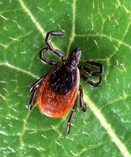
Life Cycle of Borrelia burgdorferi and Related Tick
The life cycle of Bb is related to the life cycle of the associated tick (usually Ixodes scapularis). The tick has four stages in its two-year life cycle: egg, larva, nymph and adult. The tick usually acquires the spirochete during its larval stage, when it feeds on small animals such as rodents or birds. The tick then becomes the host for the spirochete. The bacteria resides in the digestive tract of the host for its next nymph and adult stages during which it is passed on to other animals and/or humans. It has been learned that Bb may also be carried and transmitted by fleas, mosquitoes and mites.
[GRAPHIC OMITTED]
Signs and Symptoms of Infection
The first recognizable symptom following a tick bite is the development of a rash at the site within 7 to 10 days. The rash expands with an area of central clearing. Other symptoms may include low-grade fever and/or headache. The rash and early symptoms clear within 3 to 4 weeks. Multiple secondary rashes may occur following this time period. Bouts of arthritis, usually involving large joints, especially the knee, are very common. Arthritis attacks usually resolve within 3 to 4 years with or without treatment.
Early neurological complications include Bell's palsy, meningitis and encephalitis. Sub-acute symptoms may include cognitive deficits, mood and sleep disturbances, persisting for more than 10 years. One of the most common symptoms is intense fatigue. Additional symptoms may include memory loss, poor coordination, slurred speech, poor concentration, unusual depression, burning, stabbing pain, tremors, anxiety, swollen glands and tinnitus. See Chart 5.
Other conditions most commonly seen with Lyme disease include Alzheimer's disease, amyotrophic lateral sclerosis (ALS), chronic fatigue syndrome (CFS), fibromyalgia, irritable bowel syndrome, lupus, rheumatoid arthritis, scleroderma, multiple sclerosis (MS), Parkinson's disease and various autoimmune disorders. (8) See Chart 6.
Ticks may carry more than one infectious organism and, for this reason, a person infected by Bb may also be infected by other microorganisms, leading to symptoms of those diseases as well. Examples of organisms commonly occurring with Bb include Babesia microti, (25) Ehrlichia chafeensis, (26) E. equi, Mycoplasma pneumoniae, Chlamydia pneumoniae, Bartonella henselae and Rickettsia rickettsiae. The presence of multiple symptoms of several different diseases makes diagnosis and treatment of Lyme disease much more difficult. (8)
Therapy for Lyme Disease
Antibiotics
When conditions become adverse for its survival, Bb produces cysts containing the DNA defining the organism intended for future generations but surviving in a metabolically inactive state. In this state there is no cell wall generation and no way an antibiotic can damage the organism. (9)
It has been found that tetracycline can inhibit cyst formation and damage the envelope of cysts. It is also believed that bismuth compounds can enter cysts through the cyst wall. In addition, the prolonged replication rate, mentioned previously, protects the organism from cell wall damage by most antibiotics. (10) See Chart 7.
Oral Salt Therapy
Certain white blood cells (WBC) display several distinct mechanisms that may be employed for the purpose of killing invading microorganisms. One of these deserves particular attention in relation to killing the causative agent of Lyme disease, namely, the spirochete Borrelia burgdorferi.
Neutrophils (a class of WBC) contain two essentially different types of storage granules, peroxidase-positive granules and peroxidase-negative granules. Peroxidase-positive granules contain myeloperoxidase, an enzyme that uses hypochlorous acid (HOCI) in conjunction with hydrogen peroxide, providing a source of nascent (atomic) oxygen for the purpose of killing invading microorganisms. (11)
Peroxidase-negative granules contain a family of large polypeptides (11 to 19 kDa) (Dalton, the unit of molecular weight) known as the cathelicidins or, in humans, hCAP-18. A segment of this larger or precursor protein (also known as a Bacteriacidal Permeability-Increasing (BPI) protein) is proteolytically removed by the enzyme elastase found in peroxidase-positive granules. The better-known substrate of elastase is the elastic protein elastin, found in skin and other tissues requiring elasticity. By incorporating elastase inhibitors into skin creams, attempts are made to inhibit the activity of this enzyme, thereby decreasing the ageing of skin. In Lyme therapy there is an advantage (described below) to increasing the activity of this enzyme, thereby stimulating the natural antimicrobial system. These short peptides, ranging from 12 to 100 amino acids, have the ability to assemble into larger units that form pores in the membrane surrounding microorganisms, thereby increasing the permeability of those membranes. In humans, one of these antimicrobial peptides has been dubbed LL-37. (11) See Photo 10, courtesy of Blood 96 (8) 2000.
[ILLUSTRATION OMITTED]
Both of these proteins, the cathelicidin and elastase, meet in the phagocytic vacuole, the cytoplasmic chamber in which resides the phagocytized microorganism. Within this chamber, elastase removes a short peptide capable of forming a molecular pore in the surface membrane of the microorganism. The pore formed from a group of the cathelicidins allows the efflux of potassium ions from the organism, resulting in swelling and eventual lysis. (12)
Research has shown that, of all the proteins in neutrophil granules, the only protein capable of releasing the cathelicidin active peptide is elastase. (13) It has been demonstrated that the activity of elastase is enhanced by an increased salt concentration. (14) Through oral salt (12 g per day, see Chart 12), combined with large doses of vitamin C, the indirect killing ability of elastase is dramatically increased. (15)
Increasing the sodium concentration surrounding the spirochete may also facilitate cell killing by allowing sodium ions to enter the spirochete through the pore created by the antimicrobial peptide. An increased intracellular sodium concentration, combined with a decreased potassium concentration, leads to spirochete death. The exact mechanism by which the human cathelicidin LL-37 kills Bb is unknown. See Chart 8.
Colloidal Silver
It is believed that colloidal silver consisting of small clusters of silver atoms in the elemental form may be effective in eradicating Bb. If true, this action may be explained by the known ability of the Bb spirochete to bind silver, resulting in a brown/black stain. (16)
[GRAPHIC OMITTED]
Bee Venom
Bee venom is a mixture of enzymes that digest most if not all of the various kinds of biological material. Of particular value in the treatment of Lyme are the proteolytic enzymes, those that digest protein. It is believed that the proteolytic enzymes in bee venom are capable of digesting the protein coating or shell of Bb cysts. (17)
Bee venom also contains a number of potent peptides, responsible for having a strong inhibitory effect on Bb. When the spirochete is inhibited it does not multiply and is vulnerable to the host's own immune system and other medications.
Herbal Therapy
Cat's Claw
An herbal commonly used in the treatment of Lyme disease is Cat's Claw (Uncaria tomentosa), native to Peru and used for centuries by that South American culture. Traditional Cat's Claw contains chemical antagonists to the immune system known as tetracyclic oxindole alkaloids (TOASs). Some Cat's Claw products and preparations contain only the pentacyclic oxindole alkaloids (POAs. superior) for stimulating the immune system. Some are standardized for the POAs they contain. (18)
The results of research on Cat's Claw products containing POAs (Samento, commercially-available), demonstrate powerful immune system modulating and stimulating properties, along with pronounced anti-inflammatory, antioxidant, and anti-infectious effects. Cat's Claw also contains quinovic acid glycosides--compounds with strong natural antibiotic properties. (1)
Artemesia
A second herb that has been used in Lyme therapy is Artemesia annua, shown effective against Babesia, one of the more common infections accompanying Bb in Lyme patients. (19)
Bradford Research Institute/Ingles Hospital Therapy
The Bradford Research Institute (BRI)/Ingles protocols includes two antibiotics, Ciprofloxacin and Doxycycline. Also included are one or both of two new bismuth-containing compounds developed by BRI, injectible Bismacine-C and Bismacine-N. These new therapeutic agents are currently being evaluated with Lyme patients in the BRI/Ingles Hospital, Tijuana, BC, Mexico.
Oral supplementation for pain includes the following regimen:
* 4-5 g buffered Vitamin C, 2x/day
* 5 Inflazyme Forte[TM] (4000 IU Pancreatin) tablets 2x/day
* 3 Oxy-5000 Forte[TM] 2x/day
* 50 mg Magnesium Aspartate tablet one 2x/day
* Basic Elemental Minerals[TM] 2x/day
Bradford High Resolution Microscopy
As of this writing, the Bradford Research Institute/Ingles Hospital has 100% confirmation between Lyme morphology obtained utilizing the Bradford High Resolution Microscope and the Bowen fluorescent antibody test, 36 patients with positive correlation and 3 controls with negative correlation.
Rheumatoid Arthritis
In Lyme patients at the Bradford Research Institute/Ingles Hospital, it has been observed that patients with rheumatoid arthritis have dramatically improved with the Bismacine treatment along with a broad-based antimicrobial treatment (Sulfoxime[TM], Dioxychlor[R]).
In an integrative medical center, a specific treatment protocol is tailored to the individual needs, based on the concurrent assessment and diagnosis of functional pathology, organic pathology and the contributory risk factors of stress and toxicities. Based on the above assessments, an integrated treatment protocol is developed to meet the specific patient needs.
Detection of Infection
Fluorescent Antibody Test
In this test, antibodies to selected antigens on the Lyme causative organism (Bb) are chemically (covalently) attached to a chromophore, an organic chemical that fluoresces when irradiated by ultraviolet light. When the test is made, blood from the patient is mixed with the fluorescent antibody preparation. If Lyme antigens are present (during infection with Bb), the antibodies complex with and bind to the antigens present in the blood. Under ultraviolet irradiation, these molecules fluoresce, revealing the presence of Lyme antigen. This test is superior to other tests for the presence of Lyme disease and is more reliable for indicating an infection with Bb. (20)
PCR (Polymerase Chain Reaction) Test
The PCR test is very sensitive in revealing the presence of a minute amount of DNA, in this case the genetic material of Bb, indicating an infection. The test involves the amplification or multiplication of small amounts of DNA by supplying all of the requirements for the replication of DNA. The units of DNA (nucleotides) are provided, the enzyme for performing the replication is provided as well as any other required substances. If DNA from Bb is present, it will be amplified so that it may be detected by conventional means. Known nucleotide sequences of Bb are compared to those revealed by the sample. If similar, a positive identification of Bb is made. (21)
ELISA (Enzyme-Linked Immuno-Sorbant Assay)
The mechanism of the ELISA test is in some ways similar to the fluorescent antibody test described above. In the ELISA test, the antibody made by the infected person against Bb antigens as a result of infection is covalently bound to (labeled with) an enzyme, typically horseradish peroxidase. The commercially prepared and purified antigen (from Bb) as a solution is allowed to bind to the surface of small wells formed in a plate of polystyrene (commercially available). The wells are then contacted over a specified time and at a specified temperature with the enzyme-labeled antibodies (made by the patient against Bb antigens during the course of the infection and present in serum). The plate is then gently washed to remove any unbound proteins. The substrate for the enzyme is provided and the plate is allowed to develop for a specified time and at a specified temperature. If antibodies against Bb antigens are present, the covalently attached enzyme will act on the substrate provided and result in a color change. Following incubation, the color changes are read and indicate the presence of antibody, and therefore Bb antigen. This is a poor assay with marginal sensitivity for Lyme. (22)
Western Blot Test
The Western Blot test involves two electrophoretic steps in two different directions, followed by the application of antibody for specificity. In a typical procedure, serum from a Lyme patient is used as the sample protein solution in slab-gel electrophoresis on polyacrylamide gel (PAGE). The gel slabs containing the separated proteins are then employed in transverse electrophoresis using thick, carbon block (graphite) or platinum foil electrodes. Sandwiched between the gel slab and the electrodes are several layers of blotting paper soaked in electrically-conducting buffer (salts). The proteins are absorbed (blotted) onto a membrane of nitrocellulose for subsequent binding by commercially-available Bb antibody. Following the binding of specific antibody, the nitrocellulose paper is gently washed free of extraneous protein, retaining only the insoluble antigen-antibody complexes. These complexes are then stained by any of a variety of protein dyes and dried. The appearance of protein bands indicates the presence of Bb antigens in the serum. The Western Blot test has a high percentage of false negatives and should not be used to assess Lyme disease. (23)
The CDC Guidelines state that the ELISA test and the Western Blot test are plagued with false negatives and are not to be used to exclude diagnosis of Lyme disease. (27) See Chart 10.
Comparison of Detection Methods
The Centers for Disease Control (CDC) in Atlanta, Georgia, has issued guidelines for Lyme patients, advising them of a recommended protocol in attempting to establish whether Lyme disease is present or not. Doctors have been instructed by these guidelines to obtain an ELISA test first, which, under the best circumstances, identifies only 40-50% of those who actually have Lyme disease. An ELISA should NOT be used as a screening test due to the unreliable results. The guidelines then state that, if the ELISA test is positive, doctors are to perform the Western Blot test. This procedure allows many cases of Lyme disease to be missed, therefore patients are not being identified or properly treated. The CDC guidelines also state which specific bands on a nitrocellulose strip are to be used in considering a test positive. When the list of bands was developed, certain bands specific for Lyme disease were not included. When these bands are positive, they confirm exposure to the causative organism, but it is mistakenly reported to the doctor and patient as a "negative test." Many borderline tests are reported to patients as being negative and many positive tests are reported to be "false-positive" because doctors are not familiar with reading test results, nor with the multiple symptoms that can occur in a person with Lyme disease. (24,27) Charts 11, 12 and 12-A are typical Integrative Treatment Protocols for Lyme patients.
Bradford Research Institute/Ingles Hospital Protocol for Lyme Disease
The Bowen Research & Training Institute, Palm Harbor, Florida, is FDA-licensed to perform tests in which spirochetes in various forms can be detected and photographed from tissue and blood samples. They are also able to identify several strains of Babesia (25) and Ehrlichiosis. (26) This laboratory uses the fluorescent specific antibody test for detecting Bb. (20)
Charts C, D, E and F summarize the basic concepts that have been presented, including the best methods for the diagnosis and detection of Lyme disease.
Discussion
The causative organism of Lyme disease has developed the ability to not only disguise itself but to circumvent the immune system in many ways unparalleled by any other bacteria. Lyme disease has a reputation of being extremely difficult to detect and diagnose with certainty, leading many to believe they do not have the disease when, in fact, they do. The Bradford Research Institute has made significant progress in both the detection and treatment of Lyme disease, however, there is at the present time no cure or "magic bullet" for Lyme disease, implying that much additional research is greatly needed to suppress this new alarming bacterial outbreak. The Lyme epidemic has presented us with both a challenge as well as an opportunity to resolve not only Lyme, but a number of associated immunological and infectious conditions, stresses and toxicities.
Chart C Long-Standing Controversy Surrounds Lyme Disease
* There is no approved test for Lyme disease, specifically ELISA, Western Blot and PCR.
* Fate of spirochetes after entering the human system is not totally known with certainty.
* May enter B-cells, other WBC or a variety of tissues and organs.
* Professionals admit they do not know where the spirochete goes, where it hides or how it may be detected.
* NOTE: The Bradford High Resolution Microscope blood imaging has revealed the location of the spirochete and cyst form in the B-cells, eosinophils and basophils.
* Clinical evidence has revealed the associated Lyme spirochete with elevated PSA, rheumatoid arthritis, CFS, MS, fibromyalgia and diabetes.
[c]2004 BRI
Chart D -- Difficulties in Diagnosis and Detection
* The Centers for Disease Control (CDC) indicates that the number of Lyme cases may be in excess of 1.8 million.
* Other experts indicate that the figure may be in excess of 18 million in the US alone.
* Lyme disease is difficult to diagnose because it mimics many other diseases and symptoms.
* Poor detection, up to 80% false negatives using:
* ELISA (Immunological, patient's antibodies, color indicator)
* Western Blot (Electrophoresis of patient's antigens, application of specific antibody)
* PCR (Amplification of spirochete DNA, sequence comparison)
* As a result, most Lyme patients today go untreated.
[c]2004 BRI
Chart E -- Lyme Disease Out of Control
* An estimated 18 million cases in the US alone, many more worldwide.
* With poor diagnosis and treatment, there is little hope for a successful resolution.
* Available treatment modalities are only partially successful; the causative organism is masked in lymphocytes, eosinophils, basophils and tissues.
* One year ago, in the Bradford Research Institute/Ingles Hospital, Tijuana, Mexico, 1 in 20 patients had Lyme disease. Today, the incidence is in excess of 14 in 20.
Chart F Solution to Detection Problem
* The Bradford High Resolution Blood Morphology imaging of both Lyme spirochete and cyst forms have proven to be highly accurate.
* The various cyst forms are found in B-cells, eosinophils, basophils, with and without the spirochete.
* The detection of Lyme disease by the Bradford High Resolution Microscope is highly correlated with the Fluorescent Antibody Test (FDA-licensed Bowen Laboratories, Florida)
The authors wish to acknowledge Ann Marie Dixon, MBA, ND, Prof. of Medicine, Capital University of Integrative Medicine, Washington, DC, whose invaluable contribution to this manuscript is greatly appreciated.
[c] 2004 Bradford Research Institute. May be reproduced with written permission and credit.
Note: The Bradford Research Institute is seeking additional researchers to participate in The International Metabolic Research and Development Project (FDA-registered 1979), utilizing the Bradford Variable Projection Microscope. The Bradford Variable Projection Microscope blood imaging for functional assessments looks at 72 blood morphologies correlating with 111 risk factors. To qualify, the researcher must have a "scope of practice to diagnose and treat" in order to comply with the established criteria. For more information we may be contacted at: Phone: 619-429-8200, 800-227-4473, Fax: 619-429-8004, email: drbradford@americanbiologics.com. Websites: www.bradfordresearchinst.org and www.americanbiologics.com.
References
Please note: All references beginning with http://www.are internet addresses.
1. http://www.springboard4health.com/notebook/health_lyme_disease.html
2. http://www.dailymirror.lk/inside/junior/020530.html
3. http://www.intox.org/databank/documents/sodstib/ukpid80.htm
4. Sox TE, Olson CA, Binding and killing of bacteria by bismuth subsalicylate, Antimicrob Agents Chemother 1989;33:2075-82.
5. http://www.atsdr.cdc.gov/HEC/CSEM/arsenic/physiologic_effects.html
6. http://www.treedictionary.com/DICT2003/shigo/CHEM.html
7. Grier, Tom, The Complexities of Lyme Disease, from: Lyme Disease Survival Manual, 1997.
8. http://www.healingwell.com/library/lymedisease/inf02.asp
9. Alban PS, Johnson PW, Nelson DR, Serum-starvation-induced changes in protein synthesis and morphology of Borrelia burgdorferi, Microbiology 2000;146:119-27.
10. http://www.columbia-lyme.org/flatp/treatment.html
11. Borregaard N, Antibiotic Molecules: Intracellular, from: Encyclopedia of Life Sciences, 2001, Nature Publishing Group. http://immuneweb.xxmc.edu.cn/reading/innate/2.pdf
12. Wang W, Orlov D, Azimov R, et al., Mechanism of action of antimicrobial peptides: Different effects of beta-sheet and alpha-helical peptides, UCLA School of Medicine, Los Angeles, California 90095. http://meeting.biophysj.org/cgi/reprint/80/1/538/d.pdf
13. Garcia R, Gusmani L, Murgia R et al., Elastase is the only human neutrophil granule protein that alone is responsible for in vitro killing of Borrelia burgdorferi, Infect Immun 1998;66:1408-12.
14. http://www.serva.de/products/sheets/20927.shtml
15. http://lymephotos.com
16. http://www.earthbornproducts.com/dosage.htm
17. http://www.neuraltherapy.com/a_lyme_disease.asp
18. http://www.herbalremedies.com/westnile.html#1
19. http://home.pon.net/caat/lyme/artemesia.html
20. The Bowen Research & Training Institute, Connell Square, 38541 US Hwy. 19, North Palm Harbor, FL 34684
21. http://www.faseb.org/opa/bloodsupply/pcr.html
22. http://www.poultry-health.com/library/serodiss/elisa.htm
23. http://www.mcb.uct.ac.za//western.htm
24. http://www.igenex.com/lymeopt7.htm
25. http://chppm-www.apgea.army.mil/documents/FACT/18-007-0202.pdf
26. http://chppm-www.apgea.army.mil/documents/FACT/18-013-1003.pdf
27. http://www.wildernetwork.org/faq.html
28. http://www.lymenet.de/literatur/Microbiology.htm
29. Carroll MC, Lab 257: The Disturbing Story of the Government's Secret Plum Island Germ Laboratory, 2004, William Morrow Publ. Co., ISBN 0060011416. See: http://www.amazon.com.
RELATED ARTICLE: History of Lyme Disease
by Professor Robert W. Bradford and Henry W. Allen
[c] 2004 Bradford Research Institute
Correspondence:
Robert W. Bradford
Bradford Research Institute
1180 Walnut Avenue
Chula Vista, California 91911 USA
619-429-8200
drbradford@americanbiologics.com
http://www.americanbiologics.com
COPYRIGHT 2005 The Townsend Letter Group
COPYRIGHT 2005 Gale Group


 Home
Home A
A Amyotrophic lateral...