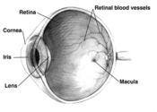
Human eye cross-sectional view. Courtesy NIH National Eye InstituteNormal vision. Courtesy NIH National Eye InstituteThe same view with age-related macular deneneration.
Find information on thousands of medical conditions and prescription drugs.

Macular degeneration

Macular degeneration is a medical condition where the light sensing cells in the macula malfunction and over time cease to work. According to the American Academy of Ophthalmology, it is the leading cause of central vision loss (blindness) in the United States today for those over the age of fifty. There are two basic types of the disease: Standard Macular Degeneration (MD) and Age Related Macular Degeneration (ARMD), with ARMD being the most common form of the condition. Macular degeneration that is not age related is most commonly caused by an inherited condition. These forms are sometimes called Juvenile macular degeneration (JMD). more...

Home
Diseases
A
B
C
D
E
F
G
H
I
J
K
L
M
Mac Ardle disease
Macroglobulinemia
Macular degeneration
Mad cow disease
Maghazaji syndrome
Mal de debarquement
Malaria
Malignant hyperthermia
Mallory-Weiss syndrome
Malouf syndrome
Mannosidosis
Marburg fever
Marfan syndrome
MASA syndrome
Mast cell disease
Mastigophobia
Mastocytosis
Mastoiditis
MAT deficiency
Maturity onset diabetes...
McArdle disease
McCune-Albright syndrome
Measles
Mediterranean fever
Megaloblastic anemia
MELAS
Meleda Disease
Melioidosis
Melkersson-Rosenthal...
Melophobia
Meniere's disease
Meningioma
Meningitis
Mental retardation
Mercury (element)
Mesothelioma
Metabolic acidosis
Metabolic disorder
Metachondromatosis
Methylmalonic acidemia
Microcephaly
Microphobia
Microphthalmia
Microscopic polyangiitis
Microsporidiosis
Microtia, meatal atresia...
Migraine
Miller-Dieker syndrome
Mitochondrial Diseases
Mitochondrial...
Mitral valve prolapse
Mobius syndrome
MODY syndrome
Moebius syndrome
Molluscum contagiosum
MOMO syndrome
Mondini Dysplasia
Mondor's disease
Monoclonal gammopathy of...
Morquio syndrome
Motor neuron disease
Motorphobia
Moyamoya disease
MPO deficiency
MR
Mucopolysaccharidosis
Mucopolysaccharidosis...
Mullerian agenesis
Multiple chemical...
Multiple endocrine...
Multiple hereditary...
Multiple myeloma
Multiple organ failure
Multiple sclerosis
Multiple system atrophy
Mumps
Muscular dystrophy
Myalgic encephalomyelitis
Myasthenia gravis
Mycetoma
Mycophobia
Mycosis fungoides
Myelitis
Myelodysplasia
Myelodysplastic syndromes
Myelofibrosis
Myeloperoxidase deficiency
Myoadenylate deaminase...
Myocarditis
Myoclonus
Myoglobinuria
Myopathy
Myopia
Myositis
Myositis ossificans
Myxedema
Myxozoa
N
O
P
Q
R
S
T
U
V
W
X
Y
Z
Medicines

In macular degeneration the final form results in missing or blurred vision in the central, reading part of vision. The outer, peripheral part of the vision remains intact.

Age related macular degeneration

ARMD is further divided into a "dry," or nonexudative, form and a "wet," or exudative, form. Eighty five to ninety percent of cases are categorized as "dry" macular degeneration where fatty tissue, known as drusen, will slowly build up behind the retina. Ten to fifteen percent of cases involve the growth of abnormal blood vessels under the retina. These cases are called "wet" macular degeneration due to the leakage of blood and other fluid from behind the retina into the eye. Wet macular degeneration usually begins as the dry form. If allowed to continue without treatment it will completely destroy the macula. Medical, photodynamic, laser photocoagulation and laser treatment of wet macular degeneration are available.

Risk factors

  • Aging: Approximately 10% of patients 66 to 74 years of age will have findings of macular degeneration. The prevalence increases to 30% in patients 75 to 85 years of age.
  • Smoking: The only environmental exposure clearly associated with macular degeneration is tobacco smoking .
  • Family history: The lifetime risk of developing late-stage macular degeneration is 50% for people who have a relative with macular degeneration vs. 12% for people who do not have relatives with macular degeneration, i.e. a four fold higher risk.
  • Macular Degeneration Gene: Complement factor H (CFH) gene has been determined to be strongly associated with a person's risk for developing macular degeneration.
  • Exposure to sunlight especially blue light.
  • Hypertension.
  • Cardiovascular Risk Factors - high cholesterol, obesity.
  • High fat intake is associated with an increased risk of macular degeneration in both women and men. Fat provides about 42 % of the calories in the average American diet. A diet that derives closer to 20-25 % of total calories from fat is probably healthier. Reducing fat intake to this level means cutting down greatly on consumption of red meats and dairy products such as milk, cheese, and butter. Eating more cold-water fish (at least twice weekly), rather than red meats and eating any type of nuts may help macular degeneration patients.(Reference: Macular degeneration Types and Risk Factors.
  • Oxidative stress: It has been proposed that age related accumulation of low molecular weight, phototoxic, pro-oxidant melanin oligomers within lysosomes in the retinal pigment epithelium (RPE) may be partly responsible for decreasing the digestive rate of photoreceptor outer rod segments (POS) by the RPE. A decrease in the digestive rate of POS has been shown to be associated with lipofuscin formation - a classic symptom of macular degeneration. (Reference: Ophthalmic Research, 2005; volume 37: pages 136-141. "Melanin aggregation and polymerization: possible implications in age related macular degeneration")
  • Race Macular degeneration is more likely to be found in whites than in blacks.

Read more at Wikipedia.org


[List your site here Free!]


Nutrient combination for macular degeneration
From Townsend Letter for Doctors and Patients, 7/1/05 by Alan R. Gaby

Ninety patients with atrophic (dry) age-related macular degeneration were randomly assigned to receive, in double-blind fashion, one of three treatments for one year: 1) lutein 10 mg/day (an amount present in approximately 60 g of spinach); 2) lutein plus a supplement containing antioxidants, vitamins, and minerals; or 3) placebo. The supplement provided daily 2,500 IU vitamin A, 15,000 IU natural beta-carotene, 1,500 mg vitamin C, 400 IU vitamin D, 500 IU vitamin E, 50 mg thiamine, 10 mg riboflavin, 70 mg vitamin B3, 50 mg pantothenic acid, 50 mg vitamin B6, 500 mcg vitamin B12, 800 mcg folic acid, 300 mcg biotin, 500 mg calcium, 300 mg magnesium, 75 mcg iodine, 25 mg zinc, 1 mg copper, 2 mg manganese, 200 mcg selenium, 200 mcg chromium, 160 mg bilberry extract (standardized to 25% anthocyanosides), 150 mg alpha-lipoic acid, 200 mg N-acetylcysteine, 100 mg quercetin, 100 mg rutin, 900 mg taurine, and several other nutrients.

The mean macular pigment optical density increased by 0.09 log unit in group 1 (p = 0.1 vs. placebo) and by 0.08 log unit in group 2 (p = 0.02), and decreased by 0.03 log unit in the placebo group. Snellen equivalent visual acuity improved by a mean of 5.4 letters (equivalent to approximately 1 line on the eye chart) in group 1 (p = 0.01 vs. baseline and vs. placebo). In group 2 the mean improvement was 3.5 letters (p = 0.04 vs. baseline; p = 0.08 vs. placebo). Contrast sensitivity improvement significantly in both active-treatment groups; the degree of improvement was greater in the group receiving combination therapy then in those receiving antioxidants alone; no significant improvement was seen in the placebo group. Active treatment was effective at all stages of age-related macular degeneration (stages II, III, & IV).

Comment: These results indicate that supplementation with lutein or lutein together with other nutrients can increase macular pigment optical density and improve visual function in patients with age-related macular degeneration, By some, but not all, measures, combination therapy was more effective than lutein alone.

Lutein is a carotenoid found primarily in spinach, corn, other vegetables, and eggs. It is a normal component of the macular pigment, where it filters the retina-damaging blue-light portion of the sun's rays. A thicker macular pigment (increased macular pigment optical density) provides greater protection than a thinner macular pigment, and lutein supplementation has been shown to increase the thickness of the macular pigment. Antioxidants such as selenium, vitamin E, vitamin C, and quercetin may protect retinal tissue against oxidative damage, and nutrients such as zinc, taurine and anthocyanosides appear to have direct effects on ocular tissues (e.g., stabilizing cell membranes or enhancing visual function).

Richer S, et al. Double-masked, placebo-controlled, randomized trial of lutein and antioxidant supplementation in the intervention of atrophic age-related macular degeneration the Veterans LAST study (Lutein Antioxidant Supplementation Trial). Optometry 2004;75(4):1-15.

COPYRIGHT 2005 The Townsend Letter Group
COPYRIGHT 2005 Gale Group

Return to Macular degeneration
Home Contact Resources Exchange Links ebay