Diagnosis of mitral valve prolapse is based on modern echocardiographic techniques which can pinpoint abnormal leaflet thickening and other related pathology.
Find information on thousands of medical conditions and prescription drugs.

Mitral valve prolapse

Mitral valve prolapse (MVP) is a heart valve condition marked by the displacement of an abnormally thickened mitral valve leaflet into the left atrium during systole. In its nonclassic form, MVP carries a low risk of complications. In severe cases of classic MVP, complications include mitral regurgitation, infective endocarditis, and — in rare circumstances — cardiac arrest usually resulting in sudden death. more...

Home
Diseases
A
B
C
D
E
F
G
H
I
J
K
L
M
Mac Ardle disease
Macroglobulinemia
Macular degeneration
Mad cow disease
Maghazaji syndrome
Mal de debarquement
Malaria
Malignant hyperthermia
Mallory-Weiss syndrome
Malouf syndrome
Mannosidosis
Marburg fever
Marfan syndrome
MASA syndrome
Mast cell disease
Mastigophobia
Mastocytosis
Mastoiditis
MAT deficiency
Maturity onset diabetes...
McArdle disease
McCune-Albright syndrome
Measles
Mediterranean fever
Megaloblastic anemia
MELAS
Meleda Disease
Melioidosis
Melkersson-Rosenthal...
Melophobia
Meniere's disease
Meningioma
Meningitis
Mental retardation
Mercury (element)
Mesothelioma
Metabolic acidosis
Metabolic disorder
Metachondromatosis
Methylmalonic acidemia
Microcephaly
Microphobia
Microphthalmia
Microscopic polyangiitis
Microsporidiosis
Microtia, meatal atresia...
Migraine
Miller-Dieker syndrome
Mitochondrial Diseases
Mitochondrial...
Mitral valve prolapse
Mobius syndrome
MODY syndrome
Moebius syndrome
Molluscum contagiosum
MOMO syndrome
Mondini Dysplasia
Mondor's disease
Monoclonal gammopathy of...
Morquio syndrome
Motor neuron disease
Motorphobia
Moyamoya disease
MPO deficiency
MR
Mucopolysaccharidosis
Mucopolysaccharidosis...
Mullerian agenesis
Multiple chemical...
Multiple endocrine...
Multiple hereditary...
Multiple myeloma
Multiple organ failure
Multiple sclerosis
Multiple system atrophy
Mumps
Muscular dystrophy
Myalgic encephalomyelitis
Myasthenia gravis
Mycetoma
Mycophobia
Mycosis fungoides
Myelitis
Myelodysplasia
Myelodysplastic syndromes
Myelofibrosis
Myeloperoxidase deficiency
Myoadenylate deaminase...
Myocarditis
Myoclonus
Myoglobinuria
Myopathy
Myopia
Myositis
Myositis ossificans
Myxedema
Myxozoa
N
O
P
Q
R
S
T
U
V
W
X
Y
Z
Medicines

Overview

The mitral valve, so named because of its resemblance to a bishop's miter, is the heart valve that prevents the backflow of blood from the left ventricle into the left atrium. It is composed of two leaflets (one anterior, one posterior) that close when the left ventricle contracts.

Each leaflet is composed of three layers of tissue: the atrialis, fibrosa, and spongiosa. Patients with classic mitral valve prolapse have excess connective tissue that thickens the spongiosa and separates collagen bundles in the fibrosa. This weakens the leaflets and adjacent tissue, resulting in increased leaflet area and elongation of the chordae tendineae. Elongation of the chordae often causes rupture, and is commonly found in the chordae tendineae attached to the posterior leaflet. Advanced lesions — also commonly involving the posterior leaflet — lead to leaflet folding, inversion, and displacement toward the left atrium.

History

For many years, mitral valve prolapse was a poorly understood anomaly associated with a wide variety of both related and seemingly unrelated signs and symptoms, including late systolic murmurs, inexplicable panic attacks, and polythelia (extra nipples). Recent studies suggest that these symptoms were incorrectly linked to MVP because the disorder was simply over-diagnosed at the time. Continuously-evolving criteria for diagnosis of MVP with echocardiography made proper diagnosis difficult, and hence many subjects without MVP were included in studies of the disorder and its prevalence. In fact, some modern studies report that as many as 55% of the population would be diagnosed with MVP if older, less reliable methods of MVP diagnosis — notably M-mode echocardiography — were used today. The term mitral valve prolapse was coined by Dr. Michael Criley in 1966 and gained acceptance over the other descriptor of "billowing" of the mitral valve (as described by Dr. Barlow).

In recent years, new criteria have been proposed as an objective measure for diagnosis of MVP using more reliable two- and three-dimensional echocardiography. The disorder has also been classified into a number of subtypes with respect to these criteria.

Subtypes

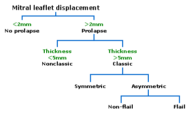
Prolapsed mitral valves are classified into several subtypes, based on leaflet thickness, concavity, and type of connection to the mitral annulus. Subtypes can be described as classic, nonclassic, symmetric, asymmetric, flail, or non-flail.

Note: all measurments below, refer to adult patients and applying them to children may be misleading

Classic vs. nonclassic

Prolapse occurs when the mitral valve leaflets are displaced more than 2 mm above the mitral annulus high points. The condition can be further divided into classic and nonclassic subtypes based on the thickness of the mitral valve leaflets: up to 5 mm is considered nonclassic, while anything beyond 5 mm is considered classic MVP.

Read more at Wikipedia.org


[List your site here Free!]


Mitral valve prolapse
From Gale Encyclopedia of Medicine, 4/6/01 by Lisa A. Papp

Definition

Mitral valve prolapse (MVP) is a ballooning of the support structures of the mitral heart valve into the left upper collection chamber of the heart.

Description

Other names for MVP include floppy valve and Barlow's syndrome. The mitral valve is located on the left side of the heart between the top chamber (left atrium) and the bottom chamber (left ventricle). The valve opens and closes according to the heartbeat and the pressure that is exerted upon it from the blood in both chambers.

The valve has supporting structures that attach to the heart muscle to help it open and close properly. When these structures weaken or lengthen abnormally, the valve may balloon into the left atrium. Sometimes this can cause the mitral valve to leak blood backward.

This condition may be inherited and occurs in approximately 10% of the population. It affects more women than men and often peaks after the age of 40.

Causes & symptoms

MVP may occur due to rheumatic heart disease but is usually found in healthy people. Changes that occur in the valve are caused by rapid multiplication of cells in the middle layer that presses on the outer layer. The outer layer weakens, causing a prolapse of the valve toward the left atrium.

Most persons do not have symptoms. Those that do may experience sharp, left-sided chest pain. Some complain of fatigue, or a pounding feeling in the chest. Others can have an irregular heart beat and even pass out. Some persons may experience difficulty breathing, ankle swelling and fluid in the lungs. Other symptoms may include anxiety, headaches, morning tiredness and constantly cold hands and feet. Death from this condition is rare.

Diagnosis

The diagnosis of MVP is based on symptoms and physical exam. During the exam, the physician may hear a click and/or heart murmur with a stethoscope.

The best diagnostic test for MVP is the echocardiogram. The test reflects sound waves through the chest wall to give two-dimensional color flow pictures of the heart, its size, position, motion, chambers, and valves. Unfortunately, during the early 1980s, this diagnosis was often made excessively from faulty echocardiographic criteria prevalent at that time.

Any person with symptoms or family history of MVP should consider having an echocardiogram. The test takes 15-20 minutes and is done in doctor's offices and hospitals. It is performed by trained technicians and is read by cardiologists. Family physicians, internists, cardiologists, and nurse practitioners can treat MVP. Echocardiograms are recommended periodically depending on the extent of valve leakage.

Treatment

Persons who experience certain types of an irregular heartbeat with MVP should be treated. Propranolol (Inderal) or other beta blockers or digoxin (Lanoxin) are often helpful. Persons who develop moderate to severe symptoms with a leaky mitral valve may require repair or replacement of the mitral valve with an artificial heart valve. Persons with MVP and a leaky valve need to protect themselves from heart or heart valve infections. Antibiotics should be taken before any surgical, dental or oral procedures according to the American Heart Association recommendations.

Other treatments include drinking lots of fluids during strenuous activity and hot weather. Water pills, caffeine and donating blood may aggravate the symptoms of MVP.

Prognosis

MVP is usually not a serious condition. However, dangerous, untreated irregular heartbeats may rarely cause sudden death. These persons should be carefully monitored.

Key Terms

Heart murmur
Sound during the heartbeat caused by a heart valve that does not close properly.
Rheumatic heart disease
A condition caused by a streptococcus infection which can result in permanent heart damage.

Further Reading

For Your Information

    Books

  • LeDoux, Denise."Acquired Valvular Heart Disease." In Cardiac Nursing, edited by Susan Woods, et al. Philadelphia.: J.B. Lippincott Co., 1995.

    Periodicals

  • McGrath, Dicey. "Mitral Valve Prolapse." American Journal of Nursing (May 1997): 40-41.

Gale Encyclopedia of Medicine. Gale Research, 1999.

Return to Mitral valve prolapse
Home Contact Resources Exchange Links ebay