Abstract
Hereditary punctate palmoplantar keratoderma (Buschke-Fischer-Brauer disease) is a rare disorder of keratinization. We describe here a 49-year-old male patient of this condition with many unusual features such as late onset of the disease in the fourth decade and nail changes (longitudinal pigmented striations, curved nails and pits in the fingernails; and nail thickening, subungual hyperkeratosis and yellowish discoloration in toenails). The patient developed histopathologically proven skin lesions typical of psoriasis 7 years after appearance of the keratoderma. This association has not been reported earlier. The patient's skin lesions cleared completely with acitretin therapy within 3 months.
Introduction
Palmoplantar keratoderma (PPKs) are uncommon disorders. They are divided into 4 categories: diffuse, focal, punctate, and the palmoplantar ectodermal dysplasias. (1) The hereditary punctate PPK (keratosis punctata palmoplantaris) is a rare form of keratoderma. Its incidence was reported as 1.17/100,000 from Croatia. (2) This entity was first described by Buschke and Fischer (3) in 1910 and Brauer (4) in 1913. Clinically, the condition usually presents with numerous pinhead-sized hyperkeratotic papules that are irregularly distributed and localized on the palms and soles. The papules may vary in size and tend to be more prominent, hyperkeratotic, and confluent on pressure points or when mechanically irritated. Both sexes are equally affected. The onset of the condition is much later than the diffuse hereditary PPK with lesions appearing in the later teens, twenties, or later. Except for some tenderness over pressure points, most patients are asymptomatic, and this disease is usually diagnosed incidentally. (5) A number of other abnormalities have been infrequently reported in association with keratosis punctata including a variety of nail dystrophies, malignancies, (especially adenocarcinoma of the colon), neurologic abnormalities like spastic paralysis, anodontia, HLA-B27-associated arthropathy, or combinations of color blindness, ulcus ventriculi or duodeni, syndactylia, and others. (6) We report here a new case of punctate PPK who developed psoriasis, an association which has not yet been reported with this condition. The patient's skin lesions responded excellently to acitretin treatment.
Case Report
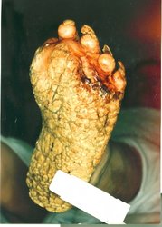
A 49-year-old male electrician from India living in Kuwait for the previous 10 years presented with multiple asymptomatic papules on his palms and soles for a duration of 7 years. Two months prior to presenting to us, he developed erythematous, scaly plaques on his hands, elbows, thighs, and buttocks. There was no history of anorexia, weight loss, constitutional, or other symptoms. He gave no history of preceding trauma, receiving immunosuppressive therapy, or exposure to arsenic. No other member in his family or neighborhood had similar lesions. The patient had received topical treatments in the form of emollients and keratolytics such as salicylic acid ointments and urea preparations in various concentrations over the last couple of years with minimal relief. No systemic treatment had been given. His general physical and systemic examinations were normal. Cutaneous examination revealed multiple, discrete, 2 to 3 mm, oval, hard, yellow, keratotic, crater-like papules located bilateral symmetrically over the palms (Figure 1a) and soles (Figure 1b). The papules were present diffusely all over the palms, including along the creases in a striking parallel rail-track pattern (Figure 1c). The papules were discrete and had not coalesced to produce plaques on the pressure points. The soles were affected more severely than the palms. There was no involvement of the sides and dorsal surface of the hands and feet with these keratotic lesions. Hyperhidrosis of the palms and soles was not present. In addition, he had erythematous, scaly plaques on his thighs (Figure 2a), gluteal regions (Figure 2b), dorsa of hands and elbows. The fingernails were curved and showed longitudinal pigmented striations and pits (Figure 3a). The toenails were thickened, had yellowish discoloration, and subungual hyperkeratosis (Figure 3b). His knees, scalp, and the rest of the body were spared. Clinical diagnosis of hereditary punctate palmoplantar keratoderma (PPK) (Brauer-Buschke-Fischer disease), punctate porokeratosis, and arsenical keratosis with psoriasis were considered.
[FIGURE 1A OMITTED]
[FIGURE 1B OMITTED]
[FIGURE 1C OMITTED]
[FIGURE 2A OMITTED]
[FIGURE 2B OMITTED]
[FIGURE 3A OMITTED]
[FIGURE 3B OMITTED]
[FIGURES 4A & 4B OMITTED]
[FIGURE 5 OMITTED]
[FIGURE 6 OMITTED]
[FIGURE 7 OMITTED]
His routine baseline investigations such as CBC, urinalysis, blood chemistry (LFT, RFT, blood sugar) and chest x-ray were normal. Ultrasound abdomen, CT scan of chest and abdomen, barium meal follow through, and upper and lower gastrointestinal endoscopies were normal. Tissue analysis for arsenic was negative. An excision biopsy of one of the papules on the left palm revealed crater like depression of the epidermis due to marked compact ortho-hyperkeratosis, an acanthotic epidermis, and mild perivascular chronic inflammation in the dermis (Figures 4a and 4b). No parakeratosis, cornoid lamella, dyskeratotic cells, or vacuolization of the epidermis was seen. The findings were consistent with the diagnosis of PPK. A biopsy from the psoriatic plaque on the left thigh revealed hyperkeratosis, parakeratosis, acanthosis, spongiform pustule formation, and upper dermal perivascular mononuclear inflammation (Figure 5). The findings were suggestive of psoriasis. The patient was treated with tablet acitretin 25 mg twice daily. Within 6 weeks the keratotic lesions had started exfoliating and by the end of 12 weeks the lesions on the palms and soles had completely cleared (Figure 6). The scaly plaques of psoriasis on the thighs, buttocks, and hands cleared even earlier within 4 weeks. The dose of acitretin was tapered to 25 mg OD for the next 3 months and then stopped. There has been no recurrence of the lesions in the last 6 months. His LFT, RFT, and serum lipid profile were monitored monthly. There was mild elevation of liver enzymes and serum triglycerides and serum cholesterol which returned to normal on stopping acitretin.
Discussion
The hereditary forms of punctate PPKs are classified into 3 types. Type I (Buschke-Fischer-Brauer disease, keratosis punctata, and keratosis papulosa) is an autosomal-dominant condition with variable penetrance. Multiple tiny, punctate keratoses are found, and there is a possible association with malignant conditions. Type II (porokeratosis punctata, palmaris, et plantaris) is also an autosomal-dominant condition with onset between ages 12 and 50. There are numerous tiny keratotic spines, which resemble the spines of a music box. Histologically, these spines correspond to columnar parakeratosis that resemble cornoid lamellae. Type III (acrokeratoelastoidosis lichenoides) is an autosomal dominant condition. There are oval or polygonal crateriform papules on the borders of the hands, feet, and wrists as well as in the center of the palms and soles. Special staining for elastin shows degeneration of elastic fibres. (1) Acquired forms of punctate PPK include arsenical keratoses, which are associated with angiosarcoma of the liver, nonmelanoma skin cancer, and bronchial adenocarcinoma; idiopathic punctate PPK, which is associated with increasing age and internal malignant conditions; idiopathic filiform porokeratotic PPK, which is associated with breast, renal, colon, and lung cancer; and punctate PPK of the palmar creases, which occurs predominantly in Afro-Caribbeans and in association with an atopic diathesis. (7) Features that support the diagnosis of Type I hereditary punctate PPK in this patient are clinical findings of numerous tiny punctate keratoses on the palms and soles and the absence of a coronoid lamella and vacuolated cells histologically.
Hereditary punctate PPK (Keratosis punctata palmoplantaris Buschke-Fischer-Brauer) is a rare genodermatosis. Several names have been given to the condition by different authors. Synonyms include keratoma dissipatum hereditarium palmare et plantare (Brauer), keratoma disseminatum, keratodermia maculosa disseminata symmetrica palmaris et plantaris (Buschke-Fischer), keratodermia punctata hereditaria, keratodermia palmoplantaris papulosa, and others. A new classification and nomenclature for keratodermas was proposed by Stevens et al in 1996. (8) The onset of hereditary punctate PPK usually occurs during puberty and adolescence between ages 12 and 30 years, but the lesions may also start to appear up to the fifth decade of life. (6)
Stevens et al (9) published a report of a family affected with punctate PPK traced for 4 generations; 49 out of 320 members were found to have the condition. Ten of the 43 adult patients (23%) developed malignancies, some of them before the age of 50 years. (9) Emmert et al (10) reported 14 families having 47 members affected with this condition. In 3 families, only one member was affected. In a majority of their patients, the lesions appeared during puberty and adolescence; however, in one patient the lesions started to appear in the sixth decade. They explained the appearance of PPK in single member-affected families as due to spontaneous mutation. Although hyperhidrosis is usually not a feature of punctate PPK, 3 of the patients in Emmert et al's report had this complaint. Our patient is also the first affected member of his family, and the lesions had a rather late onset in the fourth decade. Emmert et al (6) described another new family of punctate PPK recently in which 3 members in 3 generations, all males were affected. (6) Two cases of hereditary punctate PPK were reported recently from India. (11,12) In a large hospital-based study of hereditary keratodermas from the southern part of India, only 2 cases of idiopathic punctate PPK were reported among 31 cases detected to have keratodermas among 59,490 new dermatology patients. (13)
Nail involvement in the form of onychogryphosis, nail thickening, subungual hyperkeratosis, longitudinal fissures, and onychomadesis has been reported infrequently in punctate PPK. (14) Tosti et al (15) reported nail changes (subungual hyperkeratosis, nail pitting, onycholysis, splinter hemorrhages, and yellow-brown discoloration) in 2 patients of punctuate PPK that were clinically and histopathologically identical to those seen in psoriasis. New nail changes of onychorrhexis, onychoschizia, trachyonychia, and notching were described in the 65-year-old patient reported by Gupta et al (11) from India. Our patient also showed nail changes in the form of longitudinal pigmented striations, curved nails and pits in the fingernails; and nail thickening, subungual hyperkeratosis and yellowish discoloration in toenails. Interestingly, our patient developed psoriasis on his limbs. Although some of the nail changes infrequently described in association with punctate PPK are quite similar to psoriasis, (15) skin lesions typical of psoriasis have not been reported in such cases to date.
Keratolytic ointments, topical retinoids, and calcipotriol may not have major effects on the keratoses, except some softening. (5) Treatment with oral retinoids leads to better results, but also to more severe side effects. Baran and Juhlin (16) reported good response with etretinate in punctate PPK. Tosti et al (15) found complete clearance of skin lesions with etretinate therapy in 2 patients; however, there was no effect on nail changes. Christiansen, however, found variable response to etretinate in 9 patients of punctate PPKs. (17) Happle et al (18) observed only slight benefit in one patient of punctate PPK using this retinoid. Acitretin was reported to be rapidly effective for punctate PPK by Hesse et al. (19) Recently, a patient of punctate PPK was treated successfully with a 12-week course of continuous infusion of 5-FU at 250 mg/[m.sup.2] per day with approximately 95% clearance of the lesions. (20) A few residual lesions were noted at follow-up nearly 4 years later in this patient.
Since the affected individuals appear to be at increased risk of developing malignant conditions, a regular follow-up with thorough history taking and clinical examination is mandatory in cases of hereditary punctate PPK. Acitretin, like its prodrug etretinate, appears to be an effective and safe drug for treating such cases as shown in this patient and other reports. (15-20) As there are chances of long-term side effects with retinoids such as hyperlipidemias, careful monitoring with regular clinical follow-up and relevant laboratory investigations is warranted. The rapid and complete clearance of keratosis and psoriasis in this patient with no recurrence after stopping the drug for 6 months after follow-up is quite encouraging. The retinoids have also been used as chemoprotective agents in patients having conditions prone to develop malignancies. (21) Although the associations with malignancy to PPK has been well described in some families, the significance of the association in all cases is not known. Theoretically, retinoids may help in prevention against some of the malignancies in these patients.
Knowledge about the disease-causing gene in punctate PPK and its function might provide insights into possible skin-cancer risk. Earlier investigations excluded linkage to chromosomes 12 and 17 in some pedigrees of punctate PPKs. (22) However, Martinez-Mir et al (23) recently identified a locus for type I punctate PPK on chromosome 15q22-q24. Further genetic studies could reveal whether one entity of punctate PPK exists that is associated with increased cancer susceptibility and a broad phenotypic spectrum or whether 2 or more entities with and without cancer susceptibility exist. (6)
Our patient has probably developed punctate PPK due to a spontaneous mutation as he is the first affected member of his family. The disease appeared late in the fourth decade in him. This is probably the first report of psoriasis appearing in a patient of punctate PPK, an association difficult to explain.
References
1. Stevens HP, Leigh IM. The inherited keratodermas of palms and soles. In: Freedberg IM, Eisen AZ, Wolff K, et al, eds. Dermatology in General Medicine, 5th ed, vol 1. McGraw-Hill, New York; 1999:603-613.
2. Stanimirovic A, Kansky A, Basta-Juzbasic A, Skerlev M, Beck T. Hereditary palmoplantar keratoderma, type papulosa, in Croatia. J Am Acad Dermatol. 1993;29:435-437.
3. Buschke A, Fischer W. Keratodermia maculosa disseminata symmetrica palmaris and plantaris. Ikonographia Dermatologica. 1910;5:183-192.
4. Brauer A. Uber eine besondere Form des hereditaren Keratoms (keratoderma disseminatum hereditarium palmare et plantare). Arch Dermatol Syph. 1913;114:211-236.
5. Griffith WAD, Judge MR, Leigh IM. Disorders of Keratinization. In: Champion RH, Burton JL, Bums DA, Breathnach SM, eds. Textbook of Dermatology, 6th ed, vol 2. Blackwell Science Ltd, Oxford; 1998:1572-1573.
6. Emmert S, Kuster W, Zutt M, Hanssle H, Hallermann C, Kretschmer L, Neumann C. A new family with the rare genodermatosis keratosis punctata palmoplantaris Buschke-Fischer-Brauer. J Am Acad Dermatol. 2003;49:1166-9.
7. Asadi AK. Type I hereditary punctate keratoderma. Dermatol Online J. October 2003;9(4):38.
8. Stevens HP, Kelsell DP, Bryant SP, Bishop DT, Spurt NK, Weissenbach J, et al. Linkage of an American pedigree with palmoplantar keratoderma and malignancy (palmoplantar ectodermal dysplasia type III) to 17q24: literature survey and proposed updated classification of the keratodermas. Arch Dermatol. 1996;132:640-651.
9. Stevens HP, Kelsell DP, Leigh IM, Ostlere LS, MacDermot KD, Rustin MH. Punctate palmoplantar keratoderma and malignancy in a four-generation family. Br J Dermatol. 1996;134:720-6.
10. Emmert S, Kuster W, Hennies HC, Zutt M, Haenssle H, Kretschmer L, Neumann C. 47 patients in 14 families with the rare genodermatosis keratosis punctata palmoplantaris Buschke-Fischer-Brauer. Eur J Dermatol. 2003;13:16-20.
11. Gupta R, Mehta S, Pandhi D, Singal A. Hereditary Punctate Palmoplantar Keratoderma (PPK) (Brauer-Buschke-Fischer Syndrome). J Dermatol. 2004;31:398-402.
12. Jose S, Kamath KN, Pai GS, Pinto J. Punctate palmoplantar keratoderma. Indian J Dermatol Venereol Leprol. 2003;69:70-71.
13. Gulati S, Thappa DM, Garg BR. Hereditary palmoplantar keratodermas in South India. J Dermatol. 1997;24:765-8.
14. Stone OJ, Mullins JF. Nail changes in keratosis punctata. Arch Dermatol. 1965;92:557-8.
15. Tosti A, Morelli R, Fanti PA, Cameli N. Nail changes of punctate keratoderma: a clinical and pathological study of two patients. Acta Derm Venereol. 1993;73:66-8.
16. Baran R, Juhlin L. Keratodermia palmoplantare papuloverrucoides progressiva: successful treatment with etretinate. J Am Acad Dermatol. 1983;8:700-702.
17. Christiansen JV, Keratodermia punctata hereditaria treated with etretinate (Tigason). Acta Derm Venereol (Stockh). 1983;63:181-2.
18. Happle R, van de Kerkhof PC, Traupe H. Retinoids in disorders of keratinization: their use in adults. Dermatologica. 1987;175:107-124.
19. Hesse S, Berbis P, Privat Y. Keratodermia palmo-plantaris papulosa (Buschke-Fischer's disease): efficacy of acitretin. Br J Dermatol. 1993;128:104-5.
20. Lienemann AO, Colucci VJ, Jones MS, Trauscht JM. Treatment of palmoplantar keratoderma with continuous infusion 5-fluorouracil. Cutis. 2004;73:303-8.
21. Peck GL, Digiovanna JJ. The retinoids. In: Freedberg IM, Eisen AZ, Wolff K, et al, eds. Dermatology in General Medicine, 5th ed, vol 1. McGraw-Hill, New York; 1999:2810-2820.
22. Kelsell DP, Stevens HP, Ratnavel R, Bryant SP, Bishop DT, Leigh IM, Spurr NK. Genetic linkage studies in non-epidermolytic palmoplantar keratoderma: evidence for heterogeneity. Hum Mol Genet. June 1995;4(6):1021-5.
23. Martinez-Mir A, Zlotogorski A, Londono D, Gordon D, Grunn A, Uribe E, Horev L, Ruiz IM, Davalos NO, Alayan O, Liu J, Gilliam TC, Salas-Alanis JC, Christiano AM. Identification of a locus for type I punctate palmoplantar keratoderma on chromosome 15q22-q24. J Med Genet. 2003;40:872-8.
Address for Correspondence
Nawaf Al-Mutairi MD
PO Box 280
Farwaniya, State of Kuwait.
Telephone: 965 9370203
Fax: 965 4808167
E-mail: nalmut@usa.net
Nawaf Al-Mutairi MD FRCP(C), Arun Joshi MD, Osama Nour-Eldin MSc
Department of Dermatology, Farwaniya Hospital, Kuwait
COPYRIGHT 2005 Journal of Drugs in Dermatology, Inc.
COPYRIGHT 2005 Gale Group


