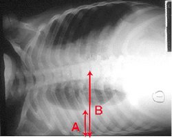
Pleural effusion Chest x-ray of a pleural effusion. The arrow A shows fluid layering in the right pleural cavity. The B arrow shows the width of the lung
Find information on thousands of medical conditions and prescription drugs.

Pleural effusion

Pleural effusion is a medical condition where fluid accumulates in the pleural cavity which surrounds the lungs, making it hard to breathe. more...

Home
Diseases
A
B
C
D
E
F
G
H
I
J
K
L
M
N
O
P
Arthritis
Arthritis
Bubonic plague
Hypokalemia
Pachydermoperiostosis
Pachygyria
Pacman syndrome
Paget's disease of bone
Paget's disease of the...
Palmoplantar Keratoderma
Pancreas divisum
Pancreatic cancer
Panhypopituitarism
Panic disorder
Panniculitis
Panophobia
Panthophobia
Papilledema
Paraganglioma
Paramyotonia congenita
Paraphilia
Paraplegia
Parapsoriasis
Parasitophobia
Parkinson's disease
Parkinson's disease
Parkinsonism
Paroxysmal nocturnal...
Patau syndrome
Patent ductus arteriosus
Pathophobia
Patterson...
Pediculosis
Pelizaeus-Merzbacher disease
Pelvic inflammatory disease
Pelvic lipomatosis
Pemphigus
Pemphigus
Pemphigus
Pendred syndrome
Periarteritis nodosa
Perinatal infections
Periodontal disease
Peripartum cardiomyopathy
Peripheral neuropathy
Peritonitis
Periventricular leukomalacia
Pernicious anemia
Perniosis
Persistent sexual arousal...
Pertussis
Pes planus
Peutz-Jeghers syndrome
Peyronie disease
Pfeiffer syndrome
Pharmacophobia
Phenylketonuria
Pheochromocytoma
Photosensitive epilepsy
Pica (disorder)
Pickardt syndrome
Pili multigemini
Pilonidal cyst
Pinta
PIRA
Pityriasis lichenoides...
Pityriasis lichenoides et...
Pityriasis rubra pilaris
Placental abruption
Pleural effusion
Pleurisy
Pleuritis
Plummer-Vinson syndrome
Pneumoconiosis
Pneumocystis jiroveci...
Pneumocystosis
Pneumonia, eosinophilic
Pneumothorax
POEMS syndrome
Poland syndrome
Poliomyelitis
Polyarteritis nodosa
Polyarthritis
Polychondritis
Polycystic kidney disease
Polycystic ovarian syndrome
Polycythemia vera
Polydactyly
Polymyalgia rheumatica
Polymyositis
Polyostotic fibrous...
Pompe's disease
Popliteal pterygium syndrome
Porencephaly
Porphyria
Porphyria cutanea tarda
Portal hypertension
Portal vein thrombosis
Post Polio syndrome
Post-traumatic stress...
Postural hypotension
Potophobia
Poxviridae disease
Prader-Willi syndrome
Precocious puberty
Preeclampsia
Premature aging
Premenstrual dysphoric...
Presbycusis
Primary biliary cirrhosis
Primary ciliary dyskinesia
Primary hyperparathyroidism
Primary lateral sclerosis
Primary progressive aphasia
Primary pulmonary...
Primary sclerosing...
Prinzmetal's variant angina
Proconvertin deficiency,...
Proctitis
Progeria
Progressive external...
Progressive multifocal...
Progressive supranuclear...
Prostatitis
Protein S deficiency
Protein-energy malnutrition
Proteus syndrome
Prune belly syndrome
Pseudocholinesterase...
Pseudogout
Pseudohermaphroditism
Pseudohypoparathyroidism
Pseudomyxoma peritonei
Pseudotumor cerebri
Pseudovaginal...
Pseudoxanthoma elasticum
Psittacosis
Psoriasis
Psychogenic polydipsia
Psychophysiologic Disorders
Pterygium
Ptosis
Pubic lice
Puerperal fever
Pulmonary alveolar...
Pulmonary hypertension
Pulmonary sequestration
Pulmonary valve stenosis
Pulmonic stenosis
Pure red cell aplasia
Purpura
Purpura, Schoenlein-Henoch
Purpura, thrombotic...
Pyelonephritis
Pyoderma gangrenosum
Pyomyositis
Pyrexiophobia
Pyrophobia
Pyropoikilocytosis
Pyrosis
Pyruvate kinase deficiency
Uveitis
Q
R
S
T
U
V
W
X
Y
Z
Medicines

Four main types of fluids can accumulate in the pleural space:

  • Serous fluid (hydrothorax)
  • Blood (hemothorax)
  • Lipid (chylothorax)
  • Pus (pyothorax or empyema)

Causes

Pleural effusion can result from reasons such as:

  • Cancer, including lung cancer or breast cancer
  • Infection such as pneumonia or tuberculosis
  • Autoimmune disease such as lupus erythematosus
  • Heart failure
  • Bleeding, often due to chest trauma (hemothorax)
  • Low oncotic pressure of the blood plasma
  • lymphatic obstruction
  • Accidental infusion of fluids

Congestive heart failure, bacterial pneumonia and lung cancer constitute the vast majority of causes in the developed countries, although tuberculosis is a common cause in the developing world.

Diagnosis

In states of excess accumulation, pleural fluid can be sampled and evaluated to determine what disease state may be causing it. This can be sampled through a thoracentesis, where a needle is inserted through the back of the chest wall and into the pleural space. The evaluation consists of:

  1. Gram stain and culture - identifies bacterial infections
  2. Cell count and differential - differentiates exudative from transudative effusions
  3. Cytology - identifies cancer cells, may also identify some infective organisms
  4. Chemical composition including protein, lactate dehydrogenase, amylase, pH and glucose - differentiates exudative from transudative effusions
  5. Other tests as suggested by the clinical situation - lipids, fungal culture, viral culture, specific immunoglobulins

Read more at Wikipedia.org


[List your site here Free!]


Medical thoracoscopy using a flexirigid thoracoscope in the diagnosis of pleural effusion of unknown cause
From CHEST, 10/1/05 by Atsuko Ishida

PURPOSE: To evaluate the efficacy of the flexirigid thoracoscope as a tool for the diagnosis in pleural effusions of unknown etiology.

METHODS: Between May 2002 and October 2004 19 patients with pleural effusion who had remained undiagnosed even after thoracentesis were performed medical thoracoscopy under local anesthesia using a flexirigid thoracoseope, a rigid thoracoscope with a bidirectional flexible tip. Patients were examined using supplemental oxygen, either in the endoscopy suite, or at the bedside in a general ward after premedicated with an intramuscular injection of hydroxyzine and pentazocine.

RESULTS: The mean age was 69 years, with a male/female ratio of 5:1. A definitive diagnosis was made in 17 of the 19 patients (89%), 6 malignant pleural mesothelioma, 5 metastatic pleural carcinoma, 4 tuberculous pleurisy, 1 empyema, and 1 uremic pleurisy. The mean examination time was 22 minutes. Complications observed were; 1 case of CO2 narcosis, 1 case of pneumothorax, and 5 cases of subcutaneous emphysema. There were no fatalities.

CONCLUSION: The flexirigid thoracoscopy under local anesthesia can be considered to be a rapid, easy, less invasive, and moreover an accurate diagnostic procedure.

CLINICAL IMPLICATIONS: It is expected to become a routine examination for pleural effusion of unknown etiology.

DISCLOSURE: Atsuko Ishida, None.

Atsuko Ishida MD * Teruomi Miyazawa MD Yuka Miyazu MD Yasuo Iwamoto MD Koji Kanoh MD Mika Zaima MD Takeo Inoue MD St. Marianna Univ. School of Medicine, Kawasaki, Japan

COPYRIGHT 2005 American College of Chest Physicians
COPYRIGHT 2005 Gale Group

Return to Pleural effusion
Home Contact Resources Exchange Links ebay