ABSTRACT
Purpose. To discuss the indications and therapeutic outcomes of synovectomy, debridement, and continuous irrigation for the treatment of pyogenic arthritis caused by intra-articular injection used in the treatment of osteoarthritis of the knee.
Methods. Records of 41 patients with infectious arthritis of the knee who presented to our hospital from 1981 were reviewed. 11 of them had a history of intraarticular injection. They underwent synovectomy, debridement, and continuous irrigation using a Salem double-lumen tube after confirmation that one side of the femorotibial joint cartilage was basically healthy.
Results. The infection was successfully treated in 9 of the 11 patients. Of these 9 patients, one died after 3 years and 2 underwent total knee arthroplasty after 3 and 8 years. The remaining 6 patients were followed up for 5 to 15 years. Five of them had deteriorating arthropathy, and the condition was unchanged in the others. Two of these 6 patients had pain while walking, and their Japanese Orthopaedic Association scores were 70. The remaining 4 had good knee function and reduction of pain, with a mean Japanese Orthopaedic Association score of 91 and a mean range of motion of 131 degrees.
Conclusion. Arthrodesis is frequently considered the treatment for osteoarthritis if the joint destruction has affected the weight-bearing surface. However, in our experience, even when inflammatory granulation develops in the cartilage surface of one side of the femorotibial joint, good results can still be obtained by synovectomy, debridement, and continuous irrigation. After the pyogenic arthritis has subsided, if osteoarthritis has advanced and bowleg has exacerbated, further treatment options are available, such as tibial resection and even joint replacement. Continuous irrigation should be considered a feasible treatment option for pyogenic arthritis.
Key words: arthritis, infectious; debridement; injections, intraarticular; irrigation; osteoarthritis, knee
INTRODUCTION
Intra-articular corticosteroid injection is frequently used for the treatment of moderate osteoarthritis (OA) of the knee in Japan. Esterhai and GeIb1 reported that although an infection caused by the intra-articular injection was rare, the likelihood of the arthrocentesis being a predisposing factor in adult septic arthritis was 23%. The articular cartilage of these pyogenic arthritic knees is already damaged. Usually, the articular cartilage of OA is affected on only one side of the weight-bearing femorotibial surface and the other side is normal.
The type of therapy, which should be selected for the treatment of the pyogenic arthritic knees, is controversial. Arthrodesis is the treatment of choice for progressive or persistent pyogenic arthritis, in which the articular cartilage is extensively destroyed. This article discusses the indications, outcomes, and follow-up observations of the use of debridement and continuous irrigation for the treatment of pyogenic arthritis that has been caused by the use of intraarticular injection.
PATIENTS AND METHODS
Since 1981, a total of 41 patients with infectious arthritis of the knee were presented to our department. 11 of the 41 patients reviewed had pyogenic arthritic knee joints caused by intra-articular injection used in the treatment of OA. The mean age of the 3 male and 8 female patients was 62 years (range, 44-76 years). Clinical characteristics of the patients with pyogenic arthritis of the knee and details of the antibiotics used are shown in Table 1.
Preoperative examination revealed that the patients had increased erythrocyte sedimentation rate (ESR) and increased C-reactive protein (CRP) levels. Six of 11 patients had symptoms of acute inflammation-fever and sudden swelling-and they experienced severe pain within 5 days after the intra-articular knee injection. Regional erythema was detected in 4 of these patients, ESR was elevated in all 6 patients, and 5 of them had increased CRP levels. The remaining 5 patients had continued chronic knee pain and elevated ESR. The CRP level was elevated slightly in 4 of these patients and a low-grade fever was detected in the other patient. Intra-articular injection had been repeatedly administered for these 5 patients, and no preceding infections had been detected around the knee or in other organs. Overall, elevation of ESR was confirmed in all 11 patients and increased CRP levels were found in 9 of them. An analysis of synovial fluid was performed in all patients, and the results showed that no crystals were present. In 6 of the 7 patients, who had the number of cells in the synovial fluid counted, the total was found to be more than 10 000/mm^sup 3^. In the remaining patient, the number of cells was 6000/mm^sup 3^, and Escherichia coli and Klebsiella pneumonie were detected from the synovial fluid. The ESR of this patient was 37 mm/h.
Arthrotomy was immediately performed in all 11 knees. After confirming that one side of the femorotibial joint cartilage was essentially healthy, synovectomy, debridement, and continuous irrigation using a Salem double-lumen tube (Nippon Sherwood, Tokyo, Japan) were performed. This treatment was based on the method described by Kawashima et al.2
Diagnosis of pyogenic arthritis was confirmed ultimately from the results of bacterial cultures or histopathological tests of the synovial membrane. The causative microorganism was detected in 9 patients.
Before inflating a pneumatic tourniquet, the affected leg was raised for 3 to 5 minutes instead of using a rubber Esmarch bandage to drive the blood out of the limb. Synovectomy and debridement were performed thoroughly, and a pulsatile washer was used intra-operatively. Two Salem double-lumen tubes (size, both over 16Fr) used for continuous irrigation were set apart to prevent a short cut. A minimal skin incision was made for the tube, which was fixed using a peritoneal button to prevent leakage of the irrigation solution. The wound was closed using monofilament nylon sutures. The suction pressure was set at 20 cm H2O. Continuous irrigation was maintained for 7 to 21 days. The tube was removed after confirming the absence of infectious agents in 3 consecutive cultures.
RESULTS
Synovectomy, debridement, and continuous irrigation were unsuccessful in improving arthritis for 2 patients, and both patients underwent arthrodesis 6 weeks later. Inflammation was successfully treated in 9 of the 11 patients. Of the 9 patients, one died after 3 years, and 2 developed OA after 3 and 8 years. These 2 patients underwent total knee arthroplasty because of joint pain and limited range of motion. The remaining 6 patients were followed up for 5 to 15 years, with 5 of them having deteriorated arthropathy, and the symptoms remaining unchanged in the other. Two of these 6 patients experienced pain while walking, their Japanese Orthopaedic Association (JOA) scores were 70. The remaining 4 had good knee function and reduction of pain, with a mean JOA score of 91 and a mean range of motion of 131 degrees (Table 2).
The patient who was followed up for the longest period in this series is reported below.
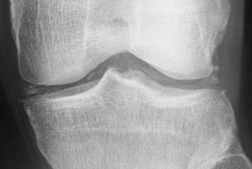
In October 1981, a 44-year-old woman was diagnosed as having OA and was treated with intraarticular steroid injections at a local clinic. The patient experienced severe pain and burning sensations one week after the intra-articular injection. In November, the patient was presented to our department. Her ESR was elevated, and radiography revealed localised bone atrophy of the medial femorotibial joint and irregular joint surfaces (Fig. 1). Arthrotomy was immediately performed because her synovial fluid was opaque.
Her synovial membrane was congested and thickened, and medial weight-bearing joint surfaces of the femur were covered by reddish-brown granulation tissue (Fig. 2). However, because the cartilage of the lateral femorotibial joint appeared normal, synovectomy, meniscus removal, debridement, and Salem double-lumen tube placement were performed. The joint was continuously irrigated with antibiotics for 14 days. A culture of synovial fluid yielded negative results, but E coli and K pneumoniae were detected from the synovial tissue fluid. Histopathological examination revealed micro-abscesses in the bone marrow.
Radiographs of the knee at 6 and 12 months postoperatively are shown in Figures 3 and 4, respectively. Bone atrophy and joint surface irregularity were improved. Her range of motion was 10 to 100 degrees during this period. The patient had occasional pain on walking during the followup period. At the final follow-up (15 years postoperatively), the patient had no pain, with range of motion of 0 to 95 degrees and a JOA score of 85. Radiograph of the knee taken at the final follow-up showed that the joint space was maintained, with a femorotibial angle of 178 degrees (Fig. 5).
DISCUSSION
The goals of treatment for pyogenic arthritis is to decrease inflammation, to preserve joint cartilage as much as possible, and to prevent the reduction of the range of motion caused by intra-articular tissue adhesion.
Early diagnosis and treatment are necessary to prevent the progress of intra-articular pyogenic inflammation.3-5 Vispo Seara et al.4 concluded that the functional outcome depended on the degenerative changes of the joint before infection, the patient's age, and the time interval between the onset of symptoms and surgical intervention. Esterhai and GeIb1 emphasised in their review that if a knee joint was affected by a sudden swelling and a severe pain, pyogenic arthritis should be considered a differential diagnosis in addition to gout, pseudogout, and apatite arthritis. Goldenberg6 and Johnson7 also discussed similar recommendations.
Patients with haematogenous pyogenic arthritis who have a history of other infections, such as pyelonephritis, usually have symptoms of fever accompanied by chills and shivering a few days before developing the arthralgia and local inflammation. However, Argen et al.8 reported that joint pain and swelling could appear suddenly without anticipating fever in patients who were on immunosuppressant medication for underlying diseases, such as malignant neoplasm or rheumatoid disease. In patients with pyogenic arthritis caused by therapeutic intraarticular injection, the condition tended to become chronic because the inflammation was suppressed by corticosteroids, although the number of pathogenic microorganisms introduced into the joint was generally small.9
Bone atrophy around the affected joint and irregularity of the subchondral bone does not appear until 7 to 10 days after the onset as seen with radiography, which can be used to reveal early pyogenic arthritis. It is helpful to compare the affected joints with the healthy ones to recognise the subtle differences. A moth-eaten appearance and joint surface destruction can be seen in the radiograph of patients with advanced pyogenic arthritis. However, these radiographic findings are non-specific in various gonarthritis.
For early diagnosis and treatment, patients with severe swelling and sudden pain should undergo an arthrocentesis to analyse the synovial fluid. Smear specimens should be analysed by Gram's stain. If a diagnosis of pyogenic arthritis is suspected, patients should undergo treatment even before the causative microorganism is identified or before results of sensitivity tests are available.
Non-surgical treatments such as antibiotic drip infusion, arthrocentesis, pus drainage, or intraarticular injection of antibiotics can be performed for the first 4 to 5 days. If joint pain and swelling persists, debridement and continuous irrigation should be performed immediately. Daniel et al.10 reported that the degree of collagen elution from joint cartilage associated with continuous irrigation of antibiotics was lower than that of systemic administration combined with pus drainage. Therefore, continuous irrigation should be performed as soon as possible.
Even when the synovial membrane is congested, synovectomy is unnecessary if the hyperplasia and thickening of synovium are mild. However, if chronic arthritis is accompanied by marked hyperplasia and thickening of synovium; alternatively, if the joint cartilage is covered by inflammatory granulation tissue, synovectomy and debridement should be performed. During the continuous irrigation, a continuous passive motion device can be used to maintain the range of motion of the affected knee joint.
Compared with conventional orthopaedic surgery, arthroscopic surgery requires a smaller incision. The advantages of better aesthetics and joint function recovery have made arthroscopy a popular technique. Arthroscopic joint irrigation, resection of pathologic synovial membrane, and adhesion detachment are also useful treatment for pyogenic arthritis.4,5,11 In a study by Skyhar and Mubarak,12 paediatric patients with pyogenic arthritis of the knee had the pyogenic joint fluid drained, and a tube was inserted to irrigate continuously through the outer sheath of the arthroscope. The low surgical invasiveness of this technique is favourable for patients with early pyogenic arthritis in which debridement is not required or for younger patients.
Arthrodesis is one of the basic surgical procedures in orthopaedic surgery. In the past, arthrodesis was performed to treat tuberculous arthritis or paralytic limb in patients with poliomyelitis. However, the use of arthrodesis has decreased13-14 because of the marked decline of these diseases and because of the advancements in artificial joint surgery.
Although various antibiotics are available today, pyogenic arthritis is still difficult to treat. Arthrodesis can be performed when arthritis becomes chronic and when the joint cartilage has been extensively destroyed. However, in our experience, even when inflammatory granulation develops on the cartilage surface on one side of the femorotibial joint, favourable results can still be obtained by synovectomy, debridement, and continuous irrigation. Even after pyogenic arthritis subsidence, if the OA advances and bowleg worsens, further treatment options are available, such as tibial osteotomy and artificial joint replacement. Continuous irrigation should be considered a feasible treatment option for pyogenic arthritis.
CONCLUSIONS
For patients with pyogenic arthritis caused by intra-articular injection, which was used to treat the OA, continuous irrigation should be performed if one side of femorotibial joint is maintained before considering arthrodesis. Therefore, early diagnosis and early continuous irrigation were crucial in treating inflammation of the knee. In our series, favourable results were obtained in 9 of 11 patients.
REFERENCES
1. Esterhai JL Jr, GeIb I. Adult septic arthritis. Orthop Clin North Am 1991;22:503-14.
2. Kawashima M, Torisu T, Kamo Y, Iwabuchi A. The treatment of pyogenic bone and joint infections by closed irrigationsuction. Clin Orthop 1980;148:240-4.
3. Studahl M, Bergman B, Kalebo P, Lindberg J. Septic arthritis of the knee: a 10-year review and long-term follow-up using a new scoring system. Scand J Infect Dis 1 994;26:85-93.
4. Vispo Seara JL, Barthel T, Schmitz H, Eulert J. Arthroscopic treatment of septic joints: prognostic factors. Arch Orthop Trauma Surg 2002;122:204-11.
5. Wirtz DC, Marth M, Miltner O, Schneider U, Zilkens KW. Septic arthritis of the knee in adults: treatment by arthroscopy or arthrotomy. Int Orthop 2001 ;25:239-41.
6. Goldenberg DL. Septic arthritis. Lancet 1 998;351:197-202.
7. Johnson MW. Acute knee effusions: a systematic approach to diagnosis. Am Fam Physician 2000;61:2391-400.
8. Argen RJ, Wilson CH Jr, Wood P. Suppurative arthritis. Clinical features of 42 cases. Arch Intern Med 1966;117:661-6.
9. Dubost JJ, Soubrier M, Sauvezie B. Pyogenic arthritis in adults. Joint Bone Spine 2000;67:11-21.
10. Daniel D, Akeson W, Amiel D, Ryder M, Boyer J. Lavage of septic joints in rabbits: effects of chondrolysis. J Bone Joint Surg Am 1976;58:393-5.
11. Stutz G, Kuster MS, Kleinstuck F, Gachter A. Arthroscopic management of septic arthritis: stages of infection and results. Knee Surg Sports Traumatol Arthrose 2000;8:270-4.
12. Skyhar MJ, Mubarak SJ. Arthroscopic treatment of septic knees in children. J Pediatr Orthop 1987;7:647-51.
13. Frymoyer JW, Hoaglund FT. The role of arthrodesis in reconstruction of the knee. Clin Orthop 1 974;101:82-92.
14. Green DP, Parkes JC 2nd, Stinchfield FE. Arthrodesis of the knee. A follow-up study. J Bone Joint Surg Am 1967;49:1065-78.
H Tsumura, S Ikeda, T Torisu
Department of Orthopedic Surgery, Faculty of Medicine, Oita University, Oita, Japan
Address correspondence and reprint requests to: Dr Hiroshi Tsumura, Associate Professor, Department of Orthopedic Surgery, Faculty of Medicine, Oita University, 1-1 ldaigaoka, Hasama-machi, Oita-gun, Oita 879-5593, Japan. E-mail: htsumura@med.oita-u.ac.jp
Copyright Western Pacific Orthopaedic Association Apr 2005
Provided by ProQuest Information and Learning Company. All rights Reserved


