Definition
Birth defects are physical abnormalities that are present at birth; they are also called congenital abnormalities. More than 3,000 have been identified.
Description
Birth defects are found in 2-3% of all newborn infants. This rate doubles in the first year, and reaches 10% by age five, as more defects become evident and can be diagnosed. Almost 20% of deaths in newborns are caused by birth defects.
Abnormalities can occur in any major organ or part of the body. Major defects are structural abnormalities that affect the way a person looks and require medical and/or surgical treatment. Minor defects are abnormalities that do not cause serious health or social problems. When multiple birth defects occur together and have a similar cause, they are called syndromes. If two or more defects tend to appear together but do not share the same cause, they are called associations.
Causes & symptoms
The specific cause of many congenital abnormalities is unknown, but several factors associated with pregnancy and delivery can increase the risk of birth defects.
Teratogens
Any substance that can causes abnormal development of the egg in the mother's womb is called a teratogen. In the first two months after conception, the developing organism is called an embryo; developmental stages from two months to birth are called fetal. Growth is rapid, and each body organ has a critical period in which it is especially sensitive to outside influences. About 7% of all congenital defects are caused by exposure to teratogens.
Drugs
Only a few drugs are known to cause birth defects, but all have the potential to cause harm. Thalidomide is known to cause defects of the arms and legs; several other types also cause problems.
- Alcohol. Drinking large amounts of alcohol while pregnant causes a cluster of defects called fetal alcohol syndrome, which include mental retardation, heart problems, and growth deficiency.
- Antibiotics. Certain antibiotics are known tetratogens. Tetracycline affects bone growth and discolors the teeth. Drugs used to treat tuberculosis can lead to hearing problems and damage to a nerve in the head (cranial). Sulfa drugs are associated with abnormally high levels of bilirubin in the newborn, which can cause death.
- Anticonvulsants. Drugs given to prevent seizures can cause serious problems in the developing fetus, including mental retardation and slow growth.
- Antipsychotic and antianxiety agents. Several drugs given for anxiety and mental illness are known to cause specific defects.
- Antineoplastic agents. Drugs given to treat cancer can cause major congenital malformations, especially central nervous system defects. They may also be harmful to the health care worker who is giving them while pregnant.
- Hormones. Male hormones may cause masculinization of a female fetus. A synthetic estrogen (DES) given in the 1940s and 1950s causes an increased risk of cancer in the adult female children of the mothers who received the drug.
- Recreational drugs. Drugs such as LSD have been associated with arm and leg abnormalities and central nervous system problems in infants. Crack cocaine has also been associated with birth defects. Since drug abusers tend to use many drugs and have poor nutrition and prenatal care, it is hard to determine the effects of individual drugs.
Chemicals
Environmental chemicals such as fungicides, food additives, and pollutants are suspected of causing birth defects, though this is difficult to prove.
Radiation
Exposure of the mother to high levels of radiation can cause small skull size (microcephaly), blindness, spina bifida, and cleft palate. How severe the defect is depends on the duration and timing of the exposure.
Infections
Three viruses are known to harm a developing baby: rubella, cytomegalovirus (CMV), and herpes simplex. Toxoplasma gondii, a parasite that can be contracted from undercooked meat, from dirt, or from handling the feces of infected cats, causes serious problems. Untreated syphilis in the mother is also harmful.
Genetic factors
A gene is a tiny, invisible unit containing information (DNA) that guides how the body forms and functions. Each individual inherits tens of thousands of genes from each parent, arranged on 46 chromosomes. Genes control all aspects of the body, how it works, and all its unique characteristics, including eye color and body size. Genes are influenced by chemicals and radiation, but sometimes changes in the genes are unexplained accidents. Each child gets half of its genes from each parent. In each pair of genes one will take precedence (dominant) over the other (recessive) in determining each trait, or characteristic. Birth defects caused by dominant inheritance include a form of dwarfism called achondroplasia; high cholesterol; Huntington's disease, a progressive nervous system disorder; Marfan syndrome, which affects connective tissue; some forms of glaucoma, and polydactyly (extra fingers or toes).
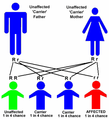
If both parents carry the same recessive gene, they have a one-in-four chance that the child will inherit the disease. Recessive diseases are severe and may lead to an early death. They include sickle cell anemia, a blood disorder that affects blacks, and Tay-Sachs disease, which causes mental retardation in people of eastern European Jewish heritage. Two recessive disorders that affect mostly whites are: cystic fibrosis, a lung and digestive disorder, and phenylketonuria (PKU), a metabolic disorder. If only one parent passes along the genes for the disorder, the normal gene received from the other parent will prevent the disease, but the child will be a carrier. Having the gene is not harmful to the carrier, but there is the 25% chance of the genetic disease showing up in the child of two carriers.
Some disorders are linked to the sex-determining chromosomes passed along by parents. Hemophilia, a condition that prevents blood from clotting, and Duchenne muscular dystrophy, which causes muscle weakness, are carried on the X chromosome. Genetic defects can also take place when the egg or sperm are forming if the mother or father passes along some faulty gene material. This is more common in older mothers. The most common defect of this kind is Down syndrome, a pattern of mental retardation and physical abnormalities, often including heart defects, caused by inheriting three copies of a chromosome rather than the normal pair.
A less understood cause of birth defects results from the interaction of genes from one or both parents plus environmental influences. These defects are thought to include:
- Cleft lip and palate, which are malformations of the mouth
- Clubfoot, ankle or foot deformities.
- Spina bifida, an open spine caused when the tube that forms the brain and spinal chord does not close properly.
- Water on the brain (hydrocephalus), which causes brain damage.
- Diabetes mellitus, an abnormality in sugar metabolism that appears later in life.
- Heart defects.
- Some forms of cancer.
A serious illness in the mother, such as an underactive thyroid, or diabetes mellitus, in which her body cannot process sugar, can also cause birth defects in the child. An abnormal amount of amniotic fluid may indicate or cause birth defects. Amniotic fluid is the liquid that surrounds and protects the unborn child in the uterus. Too little of this fluid can interfere with lung or limb development. Too much amniotic fluid can accumulate if the fetus has a disorder that interferes with swallowing.
Diagnosis
If there is a family history of birth defects or if the mother is over 35 years old, then screening tests can be done during pregnancy to gain information about the health of the baby.
If a birth defect is suspected after a baby is born, then confirmation of the diagnosis is very important. The patient's medical records and medical history may hold essential information. A careful physical examination and laboratory tests should be done. Special diagnostic tests can also provide genetic information in some cases.
- Alpha-fetoprotein test. This is a simple blood test that measure the level of a substance called alpha-fetoprotein that is associated with some major birth defects. An abnormally high or low level may indicate the need for further testing.
- Ultrasound. The use of sound waves to examine the shape, function, and age of the fetus is a common procedure. It can also detect many malformations, such as spina bifida, limb defects, and heart and kidney problems.
- Amniocentesis. This test is usually done between the 13th and 15th weeks of pregnancy. A small sample of amniotic fluid is withdrawn through a thin needle inserted into the mother's abdomen. Chromosomal analysis can rule out Down syndrome and other genetic conditions.
- Chorionic villus sampling (CVS). This test can be done as early as the ninth week of pregnancy to identify chromosome disorders and some genetic conditions. A thin needle is inserted through the abdomen or a slim tube is inserted through the vagina that takes a tiny tissue sample for testing.
Treatment
Treatment depends on the type of birth defect and how serious it is. When an abnormality has been identified before birth, then delivery can be planned at a health care facility that is prepared to offer any special care needed. Some abnormalities can be corrected with surgery. Experimental procedures have been used successfully in correcting some defects, like excessive fluid in the brain (hydrocephalus), even before the baby is born. Patients with complicated conditions usually need the help of experienced medical and educational specialists with an understanding of the disorder.
Prognosis
The prognosis for a disorder varies with the specific condition.
Prevention
Pregnant women should eat a nutritious diet. Taking folic acid supplements before and during pregnancy reduces the risk of having a baby with serious problems of the brain or spinal chord (neural tube defects). It is important to avoid any teratogen that can harm the developing baby, including alcohol and drugs. When there is a family history of congenital defects in either parent, then genetic counseling and testing can help parents plan for future children. Often, counselors can determine the risk of a genetic condition occurring and the availability of tests for it. Talking to a genetic counselor after a child is born with a defect can provide parents with information about medical management and community resources that are available.
Key Terms
- Chromosome
- One of the bodies in the cell nucleus that carries genes. There are normally 46 chromosomes in humans.
- Cleft lip and palate
- An opening in the lip, the roof of the mouth (hard palate), or the soft tissue in the back of the mouth (soft palate).
- Embryo
- The developing baby from conception to the end of the second month.
- Gene
- The The functional unit of heredity that direct all growth and development of an organism. Each human being has over 100,000 genes that determine hair color, body build, and all other traits.
- Fetus
- In humans, the developing organism from the end of the eighth week to the moment of birth.
- Neural tube defects
- A group of birth defects that affect the backbone and sometimes the spinal chord.
- Rubella
- A mild, highly contagious childhood illness caused by a virus; it is also called German measles. It causes severe birth defects if a pregnant woman is not immune and gets the illness in the first three months of pregnancy.
- Spina bifida
- One of the more common birth defects in which the backbone never closes.
- Trait
- A distinguishing feature of an individual.
- Virus
- A very small organism that causes infection and needs a living cell to reproduce.
Further Reading
For Your Information
Books
- Jones, Kenneth Lyons. "Dysmorphology." In Textbook of Pediatrics, edited by Waldo E. Nelson et al. Philadelphia: W.B. Saunders, 1996.
- Lott, Judy Wright. "Fetal Development: Environmental Influences and Critical Periods." In Comprehensive Neonatal Nursing, edited by Carol Kenner, et al. Philadelphia: W.B. Saunders, 1998.
- Van Allen, Margot I., and Judith G. Hall. "Congenital Anomalies." In Cecil Textbook of Medicine, edited by J. Claude Bennett and Fred Plum. Philadelphia: W.B. Saunders, 1996.
Periodicals
- "Down Syndrome: New Trends in Testing." Mayo Clinic Health Newsletter April 15, 1997.
Organizations
- March of Dimes Birth Defects Foundation, National Office. 1275 Mamaroneck Avenue, White Plains, New York 10605. (800) 367-6630. http://www.modimes.org.
Other
- March of Dimes. Public Health Education Information Sheets. 1994.
Gale Encyclopedia of Medicine. Gale Research, 1999.


