ABBREVIATIONS: CBC = complete blood count; IDA = iron deficiency anemia; MCHC = mean corpuscular hemoglobin concentration; MCV = mean corpuscular volume; RBC = red blood cell; TIBC = total iron binding capacity.
INDEX TERMS: Thalassemia.
Clin Lab Sci 2003;16(2):79
A 22-year-old Caucasian female clinical laboratory science student agreed to donate several anticoagulated tubes of her blood to be used for analysis in the hematology student laboratory. She was in apparent good health and had no known history of a hematological disorder. Since manual white blood cell and platelet counts were being performed that day, her blood was run on an automated instrument so that the students' values could be checked for accuracy against the instrument values. Laboratory results (Table 1) and peripheral blood smear (Figure 1) are shown.
This complete blood count (CBC) was done on an instrument which employs impedance and pulse editing technology in measuring the mean corpuscular volume (MCV). This technology has been reported to result in a 'clamped mean corpuscular hemoglobin concentration (MCHC)' which may not accurately reflect hypochromia. Hypochromic cells are more deformable and therefore present a smaller cross sectional profile as the cells pass through the aperture to be counted and sized. This results in an MCV that is reported as slightly lower than it actually is. Since the hematocrit is calculated by multiplying the MCV by the red blood cell (RBC) count, the hematocrit value will also be falsely decreased and therefore the MCHC will be somewhat overestimated. The net result is a slightly decreased sensitivity of the MCHC to the presence of hypochromia.1 However, a repeat CBC performed on a second instrument which employs pulse editing and hydrodynamic focusing reported a comparable MCHC of 32.4 g/dL. Hydrodynamic focusing has been reported to improve the clinical usefulness of the MCHC.1 On manual review, the technologist reported 1+ microcytosis and 1+ hypochromia.
QUESTIONS TO CONSIDER
1. What are the possible causes for the decreased MCV and MCH?
2. What initial tests should be performed to attempt to determine the cause of the apparent microcytosis?
3. What additional special tests might be indicated?
The findings of microcytosis and hypochromia may be associated with iron deficiency, malassemia, hemoglobin E, anemia of chronic disease, and sideroblastic anemia. Of these, the two most common causes are iron deficiency and thalassemia. Iron studies including serum iron, ferritin, and total iron binding capacity (TIBC) levels along with the red cell distribution width (RDW) can be used to help differentiate these disorders. A comparison of test results is found in Table 2. Also included in the table are results of iron studies performed on this case. It is apparent from these results that this was not a case of iron deficiency anemia (IDA). The normal results also exclude a diagnosis of sideroblastic anemia in which the percent saturation of transferrin would be elevated. The student was in good health and had no evidence of a chronic disease process. Hemoglobin electrophoresis was performed and die results were normal. From this information, a form of OC-thalassemia was suspected.
THALASSEMIA
In the normal adult the majority of hemoglobin present, approximately 97%, is hemoglobin A. Hemoglobin A is composed of two alpha globin chains and two beta globin chains ([alpha]^sub 2^[beta]^sub 2^). Other hemoglobins normally present include hemoglobin A2 ([alpha]^sub 2^[delta]^sub 2^) about 2%, and hemoglobin F ([alpha]^sub 2^[gamma]^sub 2^) about 1%. Thalassemia is an inherited disorder in which there is decreased or absent production of one or more of the globin chains. This reduced production of globin leads to the formation of microcytic, hypochromic red cells. Alpha and beta globin genes are located on chromosomes 16 and 11 respectively. Thalassemias occur in Mediterranean populations, the Middle East, parts of India and Pakistan, Southeast Asia, and Africa. However, thalassemias have also been observed in the homozygous state in persons of pure Anglo-Saxon ancestry, so ethnic origin does not preclude the diagnosis.2
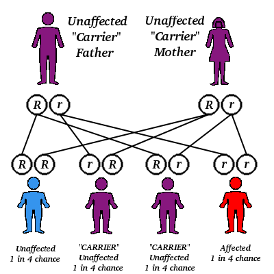
In beta thalassemia, beta chain production is reduced along with a compensatory increase in gamma and delta chain production. Over 120 beta dialassemia genes have been identified which accounts for the great diversity in the phenotypic expression of this disorder. [beta]^sup 0^ refers to a gene which produces no beta chains. [beta]^sup +^ refers to a gene which produces less than normal amounts of beta chain. Production can vary from 80% to 90% of normal to less than 10%. In the homozygous condition ([beta]^sup 0^/[beta]^sup 0^,[beta]^sup 0^/[beta]^sup +^, or [beta]^sup +^/[beta]^sup +^) anemia can be moderate, requiring minimal medical intervention, to severe, requiring regular transfusions. The majority of hemoglobin present is E Hemoglobin A is decreased or absent and hemoglobin A2 levels are slightly increased. Excess unpaired [alpha]-chains precipitate resulting in premature erythrocyte destruction and ineffective erythropoiesis. In the heterozygous condition ([beta]^sup 0^/[beta] or [beta]^sup +^/[beta]) anemia can vary from slight to moderate with slight increases in A2 and F levels.
In alpha thalassemia, alpha chain production is reduced or absent. Because there are two alpha genes (identified as [alpha]^sub 1^ and [alpha]^sub 2^) on each chromosome 16, an individual may inherit one, two, three, or four defective alpha genes. [alpha]^sup 0^refers to a deletion of both a genes on chromosome 16 (-); [alpha]^sup +^ refers to a deletion of one a gene on chromosome 16 ([alpha]-). Genotypes of [alpha]-thalassemia and the resulting phenotypic expressions are shown in Table 3.
In [alpha]-thalassemia, excess unpaired [gamma] and [beta]-chains combine to form tetramers of [gamma]^sub 4^ (Hb Barts) and [beta]^sub 4^ (Hb H). These tetramers have an extremely high oxygen affinity and are therefore poor oxygen carriers. Hb Barts and Hb H are fast moving hemoglobins and migrate past hemoglobin A on cellulose acetate at alkaline pH. Hb Barts will be present at birth but will be replaced by Hb H as the switch from [gamma]-chain to [beta]-chain synthesis occurs. In the silent carrier and thalassemia minor conditions, hemoglobin electrophoresis in the adult is normal. In hemoglobin H disease, Hb H levels vary from 5% to 40%. Hb H is somewhat unstable and can be induced to precipitate in the red cells by incubation of the blood with brilliant cresyl blue. Although demonstration of hemoglobin H inclusions is considered diagnostic of [alpha]-thalassemia trait, patients with [alpha]-/[alpha][alpha] and [alpha]-/[alpha]- genotypes usually have negative results. Patients with the -/[alpha][alpha] (trans) genotype are much more likely to test positive for hemoglobin H inclusions.3 Because hemoglobin electrophoresis is normal in [alpha]-thalassemia trait and the absence of Hb H inclusions does not rule out this disorder, diagnosis is problematic. These patients are at risk of producing homozygous (Barts hydrops fetalis) or doubly heterozygous offspring (Hb H disease) and therefore, must be identified for purposes of genetic counseling. The use of red cell parameters in differentiating between the cis and trans type has been reported. In one study, the -/[alpha][alpha] genotype was associated with lower MCVs, higher RBC counts and the presence of Hb H inclusions.3
In this case, the blood tested negative for hemoglobin H inclusions. The moderately decreased MCV, slightly elevated red cell count and negative test for Hb H inclusions were suggestive of either a [alpha]-/[alpha][alpha] (silent carrier) or [alpha]-/[alpha]- (trans) genotype.3 The father was deceased and a CBC on the mother and only sibling showed normal results.
DNA ANALYSIS
Genotyping, while expensive and time consuming, provides the definitive diagnosis. Southern blot analysis using the restriction endonucleases Bam HI and Hind III in addition to PCR assays were performed. No [alpha]^sub 2^-globin genes were detected by PCR, and Southern blot analysis confirmed the absence of the [alpha]^sub 2^-globin genes on both chromosomes. These results establish the diagnosis of [alpha]^sub 2^-thalassemia minor with a trans type genotype of [alpha]-/[alpha]-.
This form of [alpha]-thalassemia occurs with a high frequency throughout West Africa, the Mediterranean, the Middle East, and Southeast Asia.2
SUMMARY
This is a case of hypochromic, microcytic red cells in a young adult Caucasian female. It illustrates the importance of performing iron studies to confirm suspected iron deficiency anemia (IDA). Thalassemia minor is often misdiagnosed as IDA and iron therapy may be needlessly administered. Moreover, the patient will be unaware of an inherited hematological disorder which may require genetic counseling. [alpha]-thalassemia patients with the -/[alpha][alpha] (cis) genotype should be advised of the risk for producing offspring with Hemoglobin H disease (genotype -/[alpha]-).
In this case, DNA analysis confirmed the diagnosis of a trans type gene deletion [alpha]-thalassemia trait. Ancestry on the maternal side is German and French. On the paternal side the ancestry is Dutch and Scandinavian. Additionally, there was no knowledge of any family history of anemia on either the maternal or paternal side of the family. This case reaffirms that Anglo-Saxon ancestry does not preclude the diagnosis of [alpha]-thalassemia. It also supports the findings of Wang that when laboratory findings are suggestive of OC-thalassemia minor, a moderately decreased MCV, slightly elevated red cell count, and the absence of hemoglobin H inclusions is probably indicative of trans rather than cis type gene deletion [alpha]-thalassemia trait.
REFERENCES
1. Van Hove L, Schisano T, Brace L. Anemia diagnosis, classification, and monitoring using Cell-Dyn technology reviewed for the new millennium, Lab Hematol 2000;6:93-108.
2. Weatherall DJ. The thalassemias. In: Beutler E, Lichtman MA, Coller BS, Kipps TJ, editors. Williams hematology, 5th ed. New York: McGraw-Hill; 1995. p 581-615.
3. Wang C, Beganyi L, Fernandes BJ. Measurements of red cell parameters in [alpha]-thalassemia trait: correlation with the genotype. Lab Hematol 2000;6:163-6.
Angela B Foley MS CLS(NCA) is Associate Professor, Louisiana State University Health Sciences Center, New Orleans LA.
Louann W Lawrence DrPH CLSpH(NCA) is Professor and Department Head, Louisiana State University Health Sciences Center, New Orleans LA.
Address for correspondence: Angela B Foley, Department of Clinical Laboratory Sciences, School of Allied Health Professions, Louisiana State University Health Sciences Center, 1900 Gravier Street, New Orleans LA 70112, (504) 568-4276, (504) 568-6761 (fax), afoley@lsuhsc.edu
Copyright American Society for Clinical Laboratory Science Spring 2003
Provided by ProQuest Information and Learning Company. All rights Reserved


