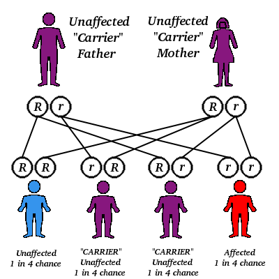
Patients with thalassemia major suffer from a qualitative defect in hemoglobin production, which causes early hemolysis, and therefore the need for repeated blood transfusions. The consequence of this hemolysis is deposition of iron in different tissues, leading to damage and dysfunction of these systems.[1,2] Iron deposition was reported to exist in autopsies of the lung. This deposition can cause lung dysfunction, most likely of a restrictive pattern. Damage to the lung can also be induced by a recurrent transfusion reaction.
At end-stage, the lungs of these patients may also be affected by cardiac damage, since many of them develop heart failure. There are conflicting reports on the state of lung function in patients with thalassemia major. While Keens et al[3] and Hoyt et al[4] reported mild decrease in expiratory flow rates, and others reported a mild restrictive pattern. The effect of transfusion on lung volume, flows, and blood gases were recently studied by Grant et al[6] in a similar group of patients who reported no ill effects. We performed a similar study evaluating the effect of blood transfusion on patients with thalassemia major. Our results seem to conflict with those of Grant and his group.[6]
Materials and Methods
Subjects
Seventeen children (8 boys, 9 girls) aged 6 to 17 years (average 11.2 years) participated in the study. The patients were 91 percent of predicted mean height for age and sex at the time of the study.[7] Six of the patients had significant growth retardation (<2 SD). All denied bronchial asthma or smoking. All were Arab residents of the Western Galilee, who received their medical care at Nahariya Hospital. Spirometry (Vitalograph) and arterial blood gases (radial artery puncture done in sitting position and measured by AVL 945) were done at baseline and 30 min following transfusion of packed cells. Lung volume and CO diffusion(DCO) were determined (Jaeger Masterlab). Diffusion was measured by the single-breath technique.[8] The cardiac status of each patient was determined by physical examination by a cardiologist, and ECG, and echo Doppler. Each patient also had a chest x-ray film. All tests were interpreted by an expert in the specific field. None of the patients received iron chelating therapy at any time.
Results
Seventeen patients were evaluated. In Table 1, a summary of their characteristics is presented. During the previous year, most of the children received transfusions of packed cells at a rate of once every three to four weeks, maintaining hemoglobin level of >10 g/dl. Six of the patients had had pneumonia of unknown cause at least once in their life.
[TABULAR DATA OMITTED]
Eight of the patients had undergone splenectomy, and 15 patients had marked hepatomegaly. The chest x-ray film, as interpreted by a certified radiologist, showed the following: six had mild increased interstitial marking (two of whom had pneumonia in the past). There was no evidence of abnormal vascular marking or ventricular enlargement. The results from the echocardiogram were as follow: patient 2 had mild left ventricular hypertrophy patient 3 had mild septal hypertrophy. Contractility was normal in all, and ECG was interpreted as normal in all the patients. Only patient 2 had evidence of cardiac malfunction on multiple tests (echo, x-ray: cardiomegaly and interstitial marking).
In Table 2, a summary of the baseline lung function is presented, as compared to their normal predicted values (for age, sex and present height).[6] The only significant abnormal function as shown in the mean value, is the diffusing capacity (corrected for anemia) which was 57 [+ or -] 12. There was also a mild decrease in forced vital capacity (85 [+ or -] 16), but all the other mean values were normal. The mean [FEV.sub.1]/VC% was particularly high (97 percent), which, together with the low diffusion capacity, might indicate a mild restrictive defect.
[TABULAR DATA OMITTED]
When analyzing each patient separately, only patients 9 and 13 had a clearly restrictive pattern. Two others (16 and 17) had spirometry results indicative of restrictive defect, but static lung volumes were not done in these patients. If one considers DCO values under 70 percent as diagnostic for abnormal diffusion capacity, eight patients out of the nine who performed the test had low diffusion capacity.
In Table 3, a summary of blood gas values is shown. Only patients 14 and 17 had arterial oxygen values below those expected at sea level for young patients (80 mm Hg).[9]
[TABULAR DATA OMITTED]
In Table 4, detailed data of the effect of transfusion on spirometry and blood gases are shown. The FVC of seven patients dropped to 10 percent or more than their pretransfusion value. Only one patient (No. 11) had a drop in [FEV.sub.1] significantly greater than the drop in FVC. The [PaO.sub.2] value in 11 patients dropped by 10 percent or more than their baseline value. Seven patients had a change of 20 percent or more, and four patients had a change greater than 40 percent, compared to their baseline value. There was no correlation between the baseline lung function, diffusion capacity, and blood gases, and the subsequent changes observed following transfusion. Nor was there any correlation between the changes in lung function and the drop seen in arterial oxygenation.
[TABULAR DATA OMITTED]
Discussion
We studied 17 patients with thalassemia major in whom detailed evaluation of pulmonary function, including spirometry, lung volumes, diffusion capacity, and blood gases were determined. Additionally, the effect of blood transfusion on spirometry and blood gases was examined in the same subjects.
Except for patient 2, none of the others had evidence of cardiac dysfunction, as determined by physical examination, ECG, chest x-ray film, and echocardiogram. One can therefore speculate that the lung function reflects primary lung pathologic condition. One major problem which arose in this study was the fact that our patients also had growth retardation, as seen in patients with long-term illness of such severity. Therefore, predicative lung volumes, as usually derived from age, height, and sex, might not be accurate, since the disease could have a different effect on the lung than on general growth.
When summarizing the baseline data, the only mean value which was significantly low, was the diffusion capacity, when compared to the predictive values.
Compared to the predictive values,[9] ten of the patients had normal (or above normal) forced vital capacity, while the remaining seven patients had low values ([is less than or equal to] 80 percent). None of the patients had an obstructive pattern, a finding which was not in agreement with that of Keens et al,[3] or Hoyt et al.[4]
The findings of reduced lung volumes and carbon monoxide diffusing capacity were in agreement with those of Cooper et al,[1] Grant et al,[6] and Grysaru et al.[10] In these studies, arterialized capillary [Po.sub.2] was mildly below the predicted value for age in the majority of the patients. Other studies[1,2] also reported hypoxia in the majority of the thalassemia patients studied. In the study of Grant el al,[6] a similar decrease in vital capacity and diffusion capacity was seen in their patients with thalassemia major. In contrast, lung volumes of our patients had a normal mean value, while theirs was reduced. This difference may reflect a different age group, since their mean age was 16.1 years, while ours was 11.2 years. The older the patient, the greater the chance of having lung damage, thus the reduced TLC. The damage could also be due to cardiac malfunction seen in patients with thalassemia.[10] Our patients, except for one, had no evidence of cardiac failure, which could be the reason for the normal TLC.
The interesting part of the study was the effect of blood transfusion on lung function seen in our patients. Most of them showed a fall in their lung volume, as reflected by the change in FVC, and an even larger number of patients had a drop in their [PaO.sub.2]. Since baseline lung function prior to blood transfusion was close to normal, one can assume that in most of the patients, the effect of blood transfusion was reversible, but in some patients, some mild restrictive lung function still remained.
Large rapid transfusion of blood has been shown to diminish vital capacity,[11] an effect which could be associated with pulmonary vascular engorgement as suggested by Gray et al.[12] Our patients developed these changes in spite of the fact that they received filtered and washed packed red blood cells, given slowly as suggested. The hypoxia seen following transfusion was also reported by Keens et al.[13] The increase in pulmonary shunting following large blood transfusion shown by Banet el al[14,15] can explain this phenomenon in our patients as well, although they did not receive large amounts of blood.
We conclude that in certain circumstances, as yet unclear, patients with thalassemia can develop hypoxemia and decrease in vital capacity following blood transfusion. Further careful studies are required to explore this unfavorable phenomenon.
References
[1] Cooper DM, Mansell Al, Weiner MA, Beadon WE, Chetty-Baktaviziam A, Reid L, et al. Lung capacity and hypoxemia in children with thalassemia major. Am Rev Respir Dis 1980; 121:639-46 [2] Witzleben CL, Wyatt JP. The effect of long survival on the pathology of thalassemia major. J Pathol Bacteriol 1961; 82:1-12 [3] Keens TG, O'Neal MH, Ortega JA, Hyman CB, Platzker ACG. Pulmonary function abnomalities in thalassemia patients on a hypertransfusion program. Pediatrics 1980; 65: 1013-17 [4] Hoyt RW, Scarpa N, Wilmott RW, Cohen A, Schwartz E. Pulmonary function abnormalities in homozygous thalassemia. J Pediatr 1986; 109:452-55 [5] Fung KP; Chow OK, Yeung SOS, Bu Yuen PM. Pulmonary function in thalassamia major. J Pediatr 1987; ill:534-37 [6] Grant GP, Mansell Al, Graziano LH, Mellins RB. The effect of transfusion on lung capacity, diffusion capacity, and arterial oxygen saturation in patients with thalassemia major. Pediatr Res 1986; 20:20-23 [7] Vaughan VC, McKay RJ, Behrman RE, Nelson WE. Growth and development. In: Nelson textbook of Pediatrics. 11th ed. Philadelphia: WB Saunders, 1979:34-35 [8] Olive CM, Foster RE, Blakemore WS, Morton JW. A standardized breath holding technique for the clinical measurement of the diffusing capacity of the lung by carbon monoxide. J Clin Invest 1957;28:1-17 [9] Polgar G, Promadhat V. Pulmonary function testing in children: techniques and standards, Philadelphia: WB Sauders, 1971 [10] Grisaru D, Rachmilewitz EA, Mosseri M, Gotsman M, Latair JS, Okon E, et al. Cardiopulmonary assessment in beta thalassemic major. Chest 1990; 98:1138-42 [11] Mollison PL. The transfusion of red cells. In:Blood transfusion in clinical medicine. 7th ed. Oxford, England: Blackwell Scientific Publications, 1983:124-130 [12] Gray BA, McCarfee DR, Sivak ED, McCurdy HT. Effect of pulmonary vascular engorgement on respiratory mechanics in the dog. J Appl Physiol 1978; 45:119-27 [13] Keens IG, O'Neal MH, Ortega JA, Hyman CB, Platzker ACG. Pulmonary function abnormalities in thalassemia patients on hypertransfusion program. Pediatrics 1980; 65:1013-17 [14] Barrett J, Tamir AH, Litwin MS. Increased pulmonary arteriovenous shunting in humans following blood transfusion. Arch Surg 1978; 113:947-50 [15] Banet J. Dawidson T. Dhurandhar HN, Miller E, Litoin MS. Pulmonary microembolism associated with massive transfusion: II. The basic pathophysiology of its pulmonary effects. Ann Surg 1975; 182:56-61
COPYRIGHT 1992 American College of Chest Physicians
COPYRIGHT 2004 Gale Group


